Electron Microscopy
Electron microscopy (EM) is a method for obtaining high-resolution images of biological and non-biological specimens. These instruments detail the structures of cells, organelles and macromolecular complexes.
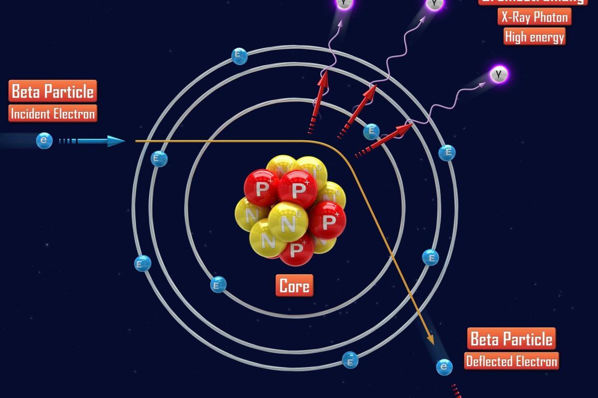
The high resolution of images results from electrons’ wavelength and is the foundation of the illuminating radiation. Electron microscopy is used in conjunction with other methods, for example, thin sectioning, immuno-labelling and staining) to answer particular questions. EM images provide essential information on the structural basis of cell function and disease. The two main types of electron microscopes include Transmission Electron Microscopy (TEM) and Scanning Electroarestransmission electron microscopyctron microscanning electron microscopye structures, such as cell sections and molecules, by which electrons can move to generate a projection image. The TEM is analogous in several ways to the traditional light microscope. For example, TEM can be used to image the inside of cells, the structure of protein molecules, the role of molecules in infections and the set-up of protein molecules in cell membranes.
Scanning electron microscopy depends upon the emission of secondary electrons from the surface of a specimen. It can produce detailed images of the areas of cells and entire organisms that are not feasible by TEM. The SEM can be used for particle counting, size dedication, and procedure control. It is called a scanning electron microscope since the image is created by scanning a concentrated electron beam onto the top of the specimen in a raster design.
The primary electron beam interacts with the atoms close to the surface and results in the emission of particles. These particles are collected with several detectors, and their relative quantity is translated into photons on the cathode ray tube (CRT). As the size of the raster at the specimen is much smaller than the viewing display of the CRT, the ultimate picture can be a magnified image of the specimen. SEMs with secondary, backscatter and X-ray detectors can review specimens’ topography and atomic composition.
home » electron microscopy
 By
By