PET Imaging
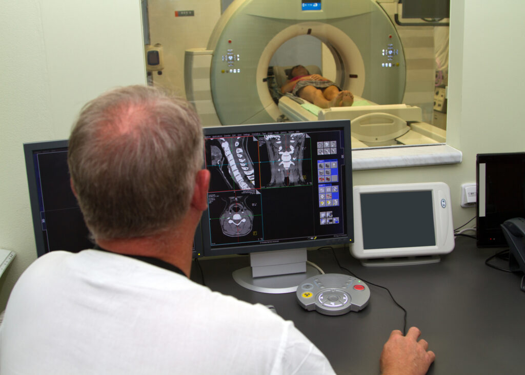
PET (Positron Emission Tomography) is a nuclear medicine imaging modality used to visualize the structures within the human body. In most cases, a PET scan is performed in conjunction with a computed tomography (CT) scan, a process known as hybrid imaging.
Consequently, these noninvasive imaging procedures are rapid and sensitive to anatomical and biological changes in the body and provide essential information about how a particular disease behaves in patients, for example, in staging, restaging, and assessing cancer treatment response.
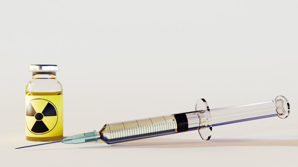
During the PET imaging procedure, the patient is injected with a low radiopharmaceutical (radiotracer) dose such as 18FDG, Na18F, 68Ga-PSMA and 68Ga-DOTATATE. However, conventional CT and MRI (magnetic resonance imaging) use X-rays to obtain 3D anatomical images. An injected radiation dose into the human body distributes to specific areas distributed close metabolism when using 18FDG to identify absorptivity used in the body.
PET imaging also enables the determination of a tumour’s metabolism rate to see if it is still active. A further hybrid uses PET and MRI to facilitate improvement in the visualisation of specific structures, for example, in the brain and pelvic organs.
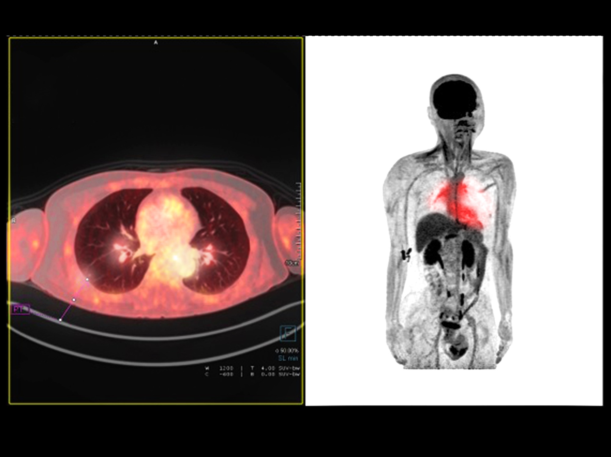
In another situation regarding lung cancer diagnosis, it is paramount for the oncologist to determine if the patient requires curative surgery and chemotherapy or only palliative chemotherapy. Therefore, to enable this decision, using 18FDG radiotracer in a PET-CT scan will allow the oncologist to verify if the lung cancer has spread to a particular lymph node.
The essence of PET imaging is to evaluate metabolic changes that precede anatomical changes, which MRI observes. Consequently, positron emission tomography imaging detects tumours early and assesses dementia and seizures. It also observes the functions of the heart and evaluates inflammatory diseases.
home » PET imaging » Page 3
 By
By