Technetium-99m Radiopharmaceuticals in Advanced Medical Imaging
 By
By
Technetium-99m, discovered in 1937, transformed medical imaging with its versatile and safe diagnostic applications.
By
Technetium-99m, discovered in 1937, transformed medical imaging with its versatile and safe diagnostic applications.
 By
By
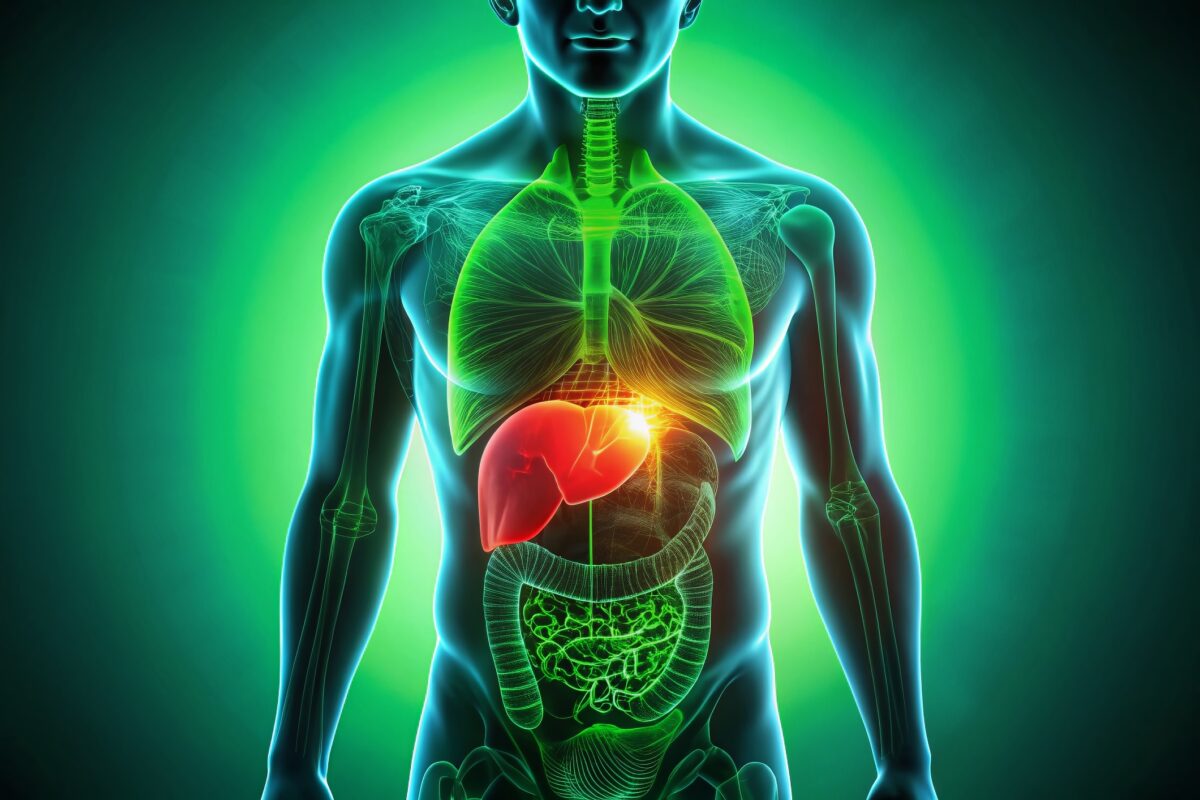
Technetium-99m sulfur colloid provides critical diagnostics for liver, spleen, and bone marrow using targeted nuclear imaging.
 By
By
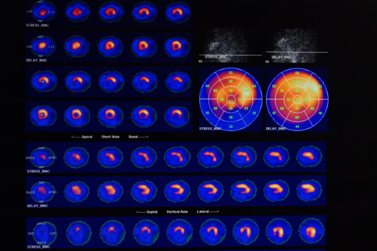
Technetium-99m Sestamibi revolutionises cardiac care by precisely mapping blood flow in diagnosing coronary artery disease.
By
Technescan, utilising Technetium-99m oxidronate, revolutionises bone imaging by highlighting metabolic activity with high sensitivity and precision.
By
Technetium-99m medronate is vital in nuclear medicine for precise bone imaging and detecting osteogenesis abnormalities.
By
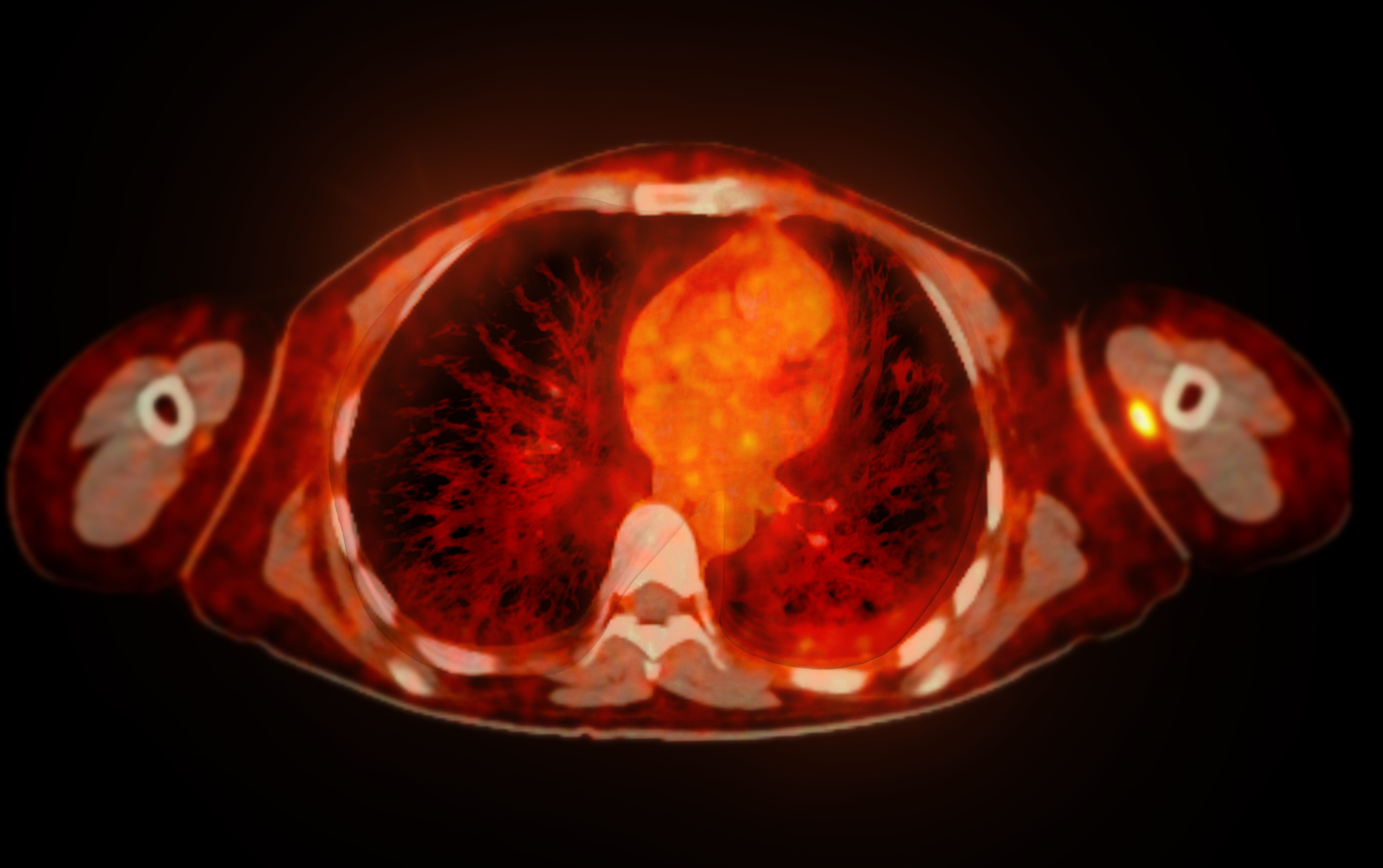
Technetium-99m Macroaggregated Albumin serves a key role in assessing pulmonary vascular integrity, supporting targeted therapeutic measures.
By
Technetium-99m exametazime (Ceretec) is a pivotal radiopharmaceutical in nuclear medicine, revolutionising brain imaging with high precision and reliability.
By
Technetium-99m bicisate (Neurolite) facilitates stroke diagnosis by enabling cerebral perfusion imaging, aiding in locating affected brain regions.
 By
By
Bone imaging is an essential diagnostic tool for detecting bone diseases, injuries, and disorders.