
Common Causes of Leg Pain and When to See a Doctor
 By
By
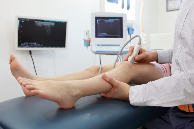
Understanding leg pain can help you decide when to consult a doctor. Discover patterns and symptoms that matter.
By
Understanding leg pain can help you decide when to consult a doctor. Discover patterns and symptoms that matter.
 By
By
Learn about effective knee pain treatment methods focusing on less invasive solutions for better joint function and comfort.
 By
By
Discover how digital optical comparators revolutionize dimensional inspection for orthopedic implants and improve patient safety.
 By
By
Learn how bone regeneration biomaterials are revolutionizing implant dentistry for patients with complex anatomical challenges.
 By
By
Learn if indigestion can cause back pain and identify symptoms to distinguish it from other back issues. Image for illustration only. Person depicted is a model.
 By
By
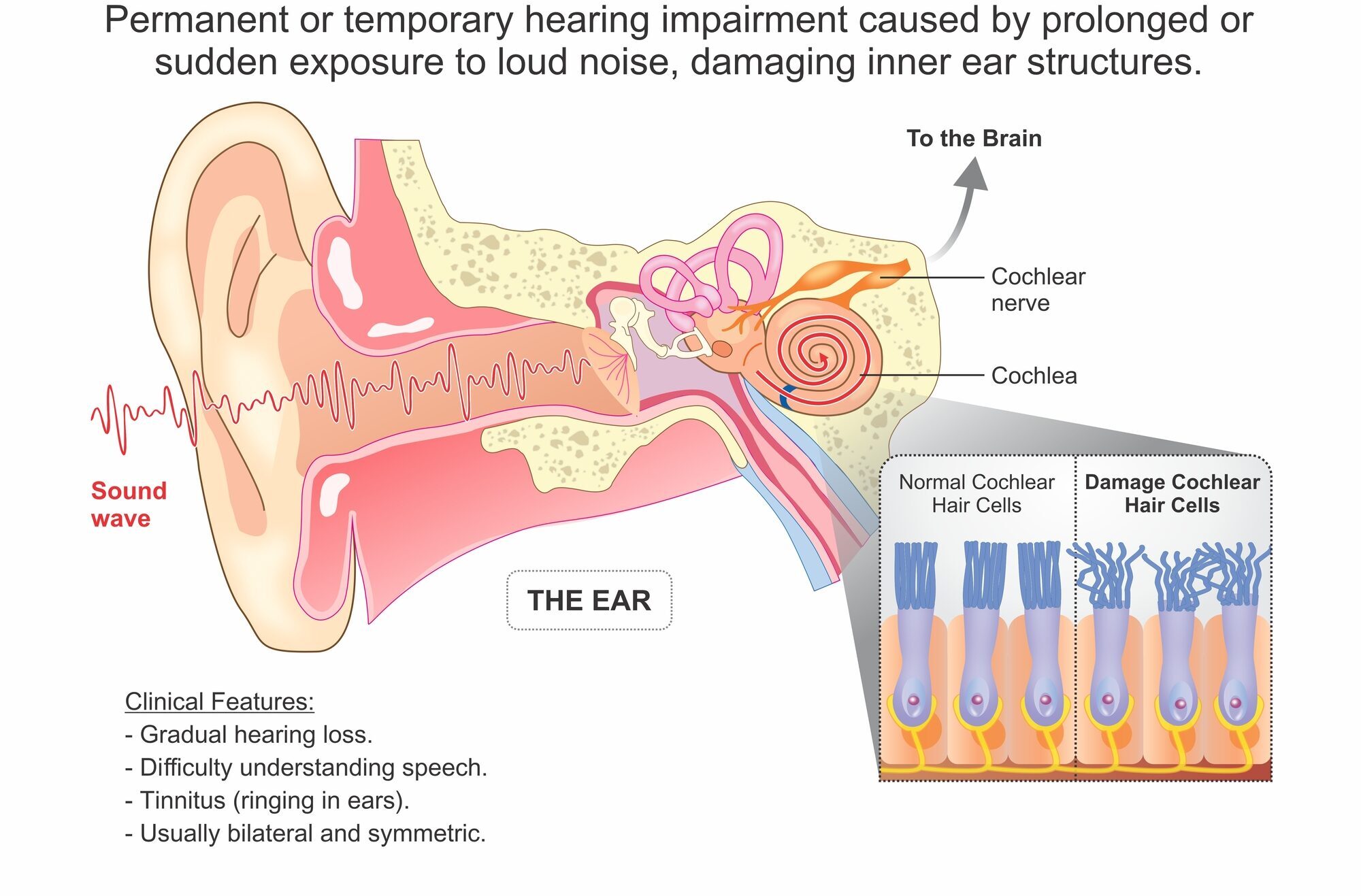
Sudden hearing loss in one ear can be alarming. Understand the symptoms and seek timely treatment for better recovery.
 By
By
Learn how wound care certification can improve your ability to handle diabetic foot ulcers and pressure injuries confidently.
 By
By
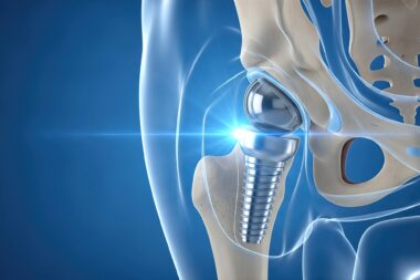
Discover how hip replacement imaging has advanced, improving diagnosis and surgical planning for better mobility outcomes.
 By
By
Digestive issues are common among runners. Find out how to address these problems for a more enjoyable running experience.
 By
By
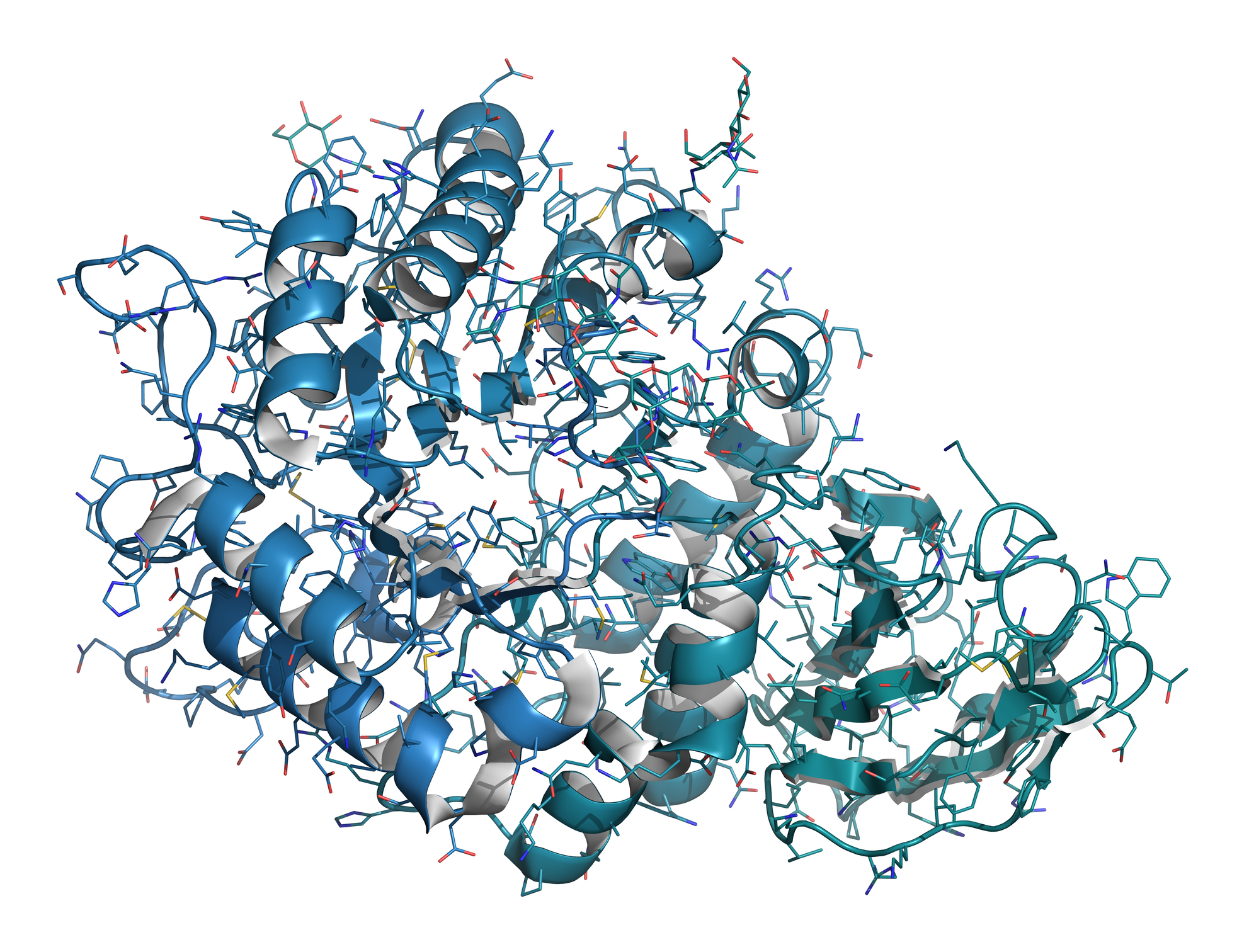
Learn how Follistatin-344 research is reshaping our understanding of cellular growth modulation and ligand binding mechanisms.
 By
By
Knee health is essential for mobility, preventing pain, strengthening joints, improving flexibility, and maintaining an active, injury-free lifestyle daily.
 By
By
Yttrium-90 Besilesomab targets CD66-expressing cells, providing precise beta radiation therapy for systemic amyloid light chain amyloidosis.
 By
By
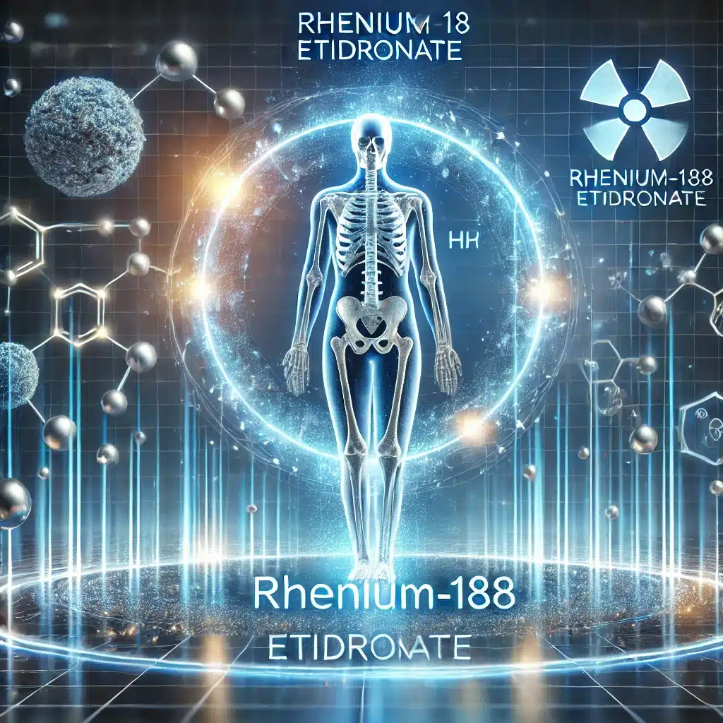
Rhenium-188 Etidronate effectively targets metastatic bone lesions, delivering therapeutic beta radiation while reducing associated pain.
 By
By
32P-Sodium Phosphate effectively reduces metastatic bone pain while managing Polycythaemia Vera, enhancing patient quality of life.
 By
By
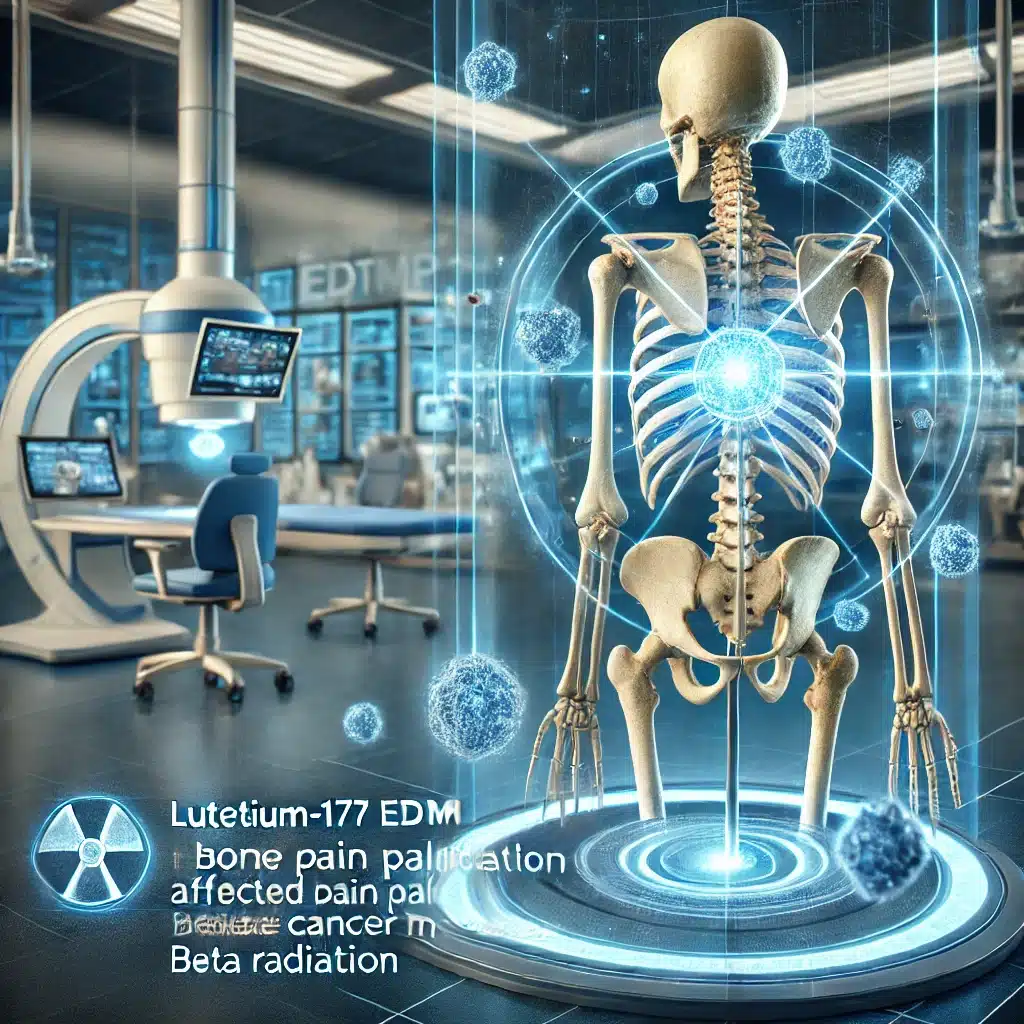
Lutetium-177 EDTMP provides a cost-effective, accessible, and efficacious alternative for bone pain palliation in metastatic cancer patients.
 By
By
Lutetium-177 DOTAZOL offers a dual-function approach, combining targeted radiotherapy and bone modulation, enhancing treatment for prostate cancer-related bone metastases.
 By
By
Bone densitometry, specifically DEXA scanning, is essential for diagnosing osteoporosis, assessing fracture risk, and monitoring treatment efficacy in patients.
 By
By
Holmium-166 Phytate, initially developed for chronic synovitis, has shown significant promise in phase I/II clinical trials.
 By
By
The innovative 169Er-Erbium Citrate therapy precisely targets small joints, significantly easing arthritis with minimal side effects.
 By
By
Technetium-99m pyrophosphate is pivotal for diagnosing bone diseases and cardiac amyloidosis through targeted radiopharmaceutical imaging.
By
Technescan, utilising Technetium-99m oxidronate, revolutionises bone imaging by highlighting metabolic activity with high sensitivity and precision.
By
Fluorine-18 Sodium Fluoride (18F-NaF) is an effective, safe diagnostic radiopharmaceutical, revolutionising bone imaging and detecting skeletal abnormalities with high accuracy.
 By
By
Bone imaging is an essential diagnostic tool for detecting bone diseases, injuries, and disorders.