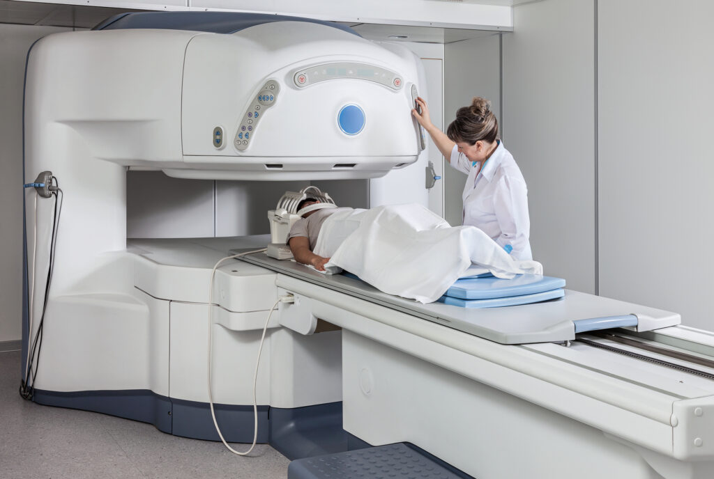
Alzheimer’s disease stems from genetic mutations and lifestyle factors, leading to amyloid plaques and cognitive decline.
Alzheimer’s Disease: Genetic, Environmental, and Lifestyle Factors
Alzheimer’s disease (AD) is a complex and progressive neurodegenerative disorder that is the most common cause of dementia. The exact cause of AD is not fully understood and is believed to result from multiple factors, including genetic, environmental, and lifestyle influences that affect the brain over time.
Genetically, Alzheimer’s disease can be split into familial (early-onset) and sporadic (late-onset) forms. Early-onset AD is rare, accounting for less than 5% of cases, and is caused by mutations in one of three genes: the amyloid precursor protein (APP), presenilin 1 (PSEN1), or presenilin 2 (PSEN2). These mutations lead to the abnormal production of amyloid-beta peptide, which aggregates into plaques that are toxic to neurons. Late-onset AD is far more common and has a more complex genetic component. The apolipoprotein E (APOE) gene, specifically the APOE ε4 allele, is known to increase the risk of AD. Still, it is not deterministic, meaning not everyone with the allele will develop the disease.
Environmental factors and lifestyle are also implicated in the risk of developing Alzheimer’s disease. Cardiovascular health is a significant factor; conditions such as hypertension, diabetes, obesity, and hypercholesterolemia can increase the risk of AD, likely due to their effects on cerebral blood flow and brain metabolism. Head injuries and exposure to certain toxins or pollutants have also been associated with an increased risk of AD.
Lifestyle choices may influence the risk of developing Alzheimer’s. A diet rich in fruits, vegetables, and fatty fish, regular physical activity, social engagement, and cognitive stimulation have all been associated with a reduced risk of AD. Conversely, a diet high in saturated fats, smoking, excessive alcohol consumption, and a sedentary lifestyle may increase the risk.
On the pathophysiological level, Alzheimer’s disease is characterised by two key abnormalities: amyloid plaques and neurofibrillary tangles. Amyloid plaques are accumulations of amyloid-beta peptides outside neurons, and neurofibrillary tangles are twisted fibres of a protein called tau inside neurons. The exact relationship between these abnormalities and the progression of AD is a subject of ongoing research, but it is believed that they disrupt the communication between neurons and trigger inflammatory processes and neuronal death, leading to the symptoms of dementia.
An additional factor is the loss of synaptic connections and the death of neurons in regions of the brain that are critical for memory and cognitive function, such as the hippocampus. The brain also experiences a reduction in neurotransmitters, chemicals that are essential for communication between neurons, particularly acetylcholine, which is involved in memory and learning.
Amyloid PET Imaging: Illuminating Alzheimer’s Pathology and Advancing Diagnostic Precision
Amyloid PET (positron emission tomography) imaging is a nuclear medicine technique used to visualise amyloid plaques in the brain, which are hallmarks of Alzheimer’s disease (AD) and other types of dementia. This imaging tool has become increasingly important in both research settings and clinical practice, as it provides a non-invasive method to detect amyloid deposition in vivo.
The process involves the administration of a radioactive tracer that binds specifically to amyloid plaques. The most commonly used tracers are carbon-11 labelled Pittsburgh Compound B (PiB), and fluorine-18 labelled tracers such as flutemetamol, florbetapir, and florbetaben. Once the tracer is administered, it crosses the blood-brain barrier and binds to amyloid plaques, if present. The PET scanner then detects the radioactive signal and generates images that reflect the brain’s distribution and density of amyloid plaques.
The ability to visualise amyloid plaques has significant implications for diagnosing and managing AD. Amyloid PET imaging can help differentiate AD from other types of dementia, as not all dementias involve amyloid plaque deposition. It is also used in clinical trials to select participants with evidence of amyloid pathology and assess the efficacy of treatments to reduce amyloid burden.
Although it has benefits, amyloid PET imaging has limitations. The presence of amyloid plaques does not necessarily correlate with the severity of dementia symptoms, meaning that a positive amyloid PET scan does not always indicate that a person will progress to AD. Additionally, the cost of PET scans and the limited availability of tracers can be prohibitive. There are also concerns about the interpretation of PET images, as the distinction between pathological and normal ageing-related amyloid deposition can be challenging.
Amyloid PET Imaging Breakthroughs
In terms of research, amyloid PET imaging has facilitated a deeper understanding of the temporal sequence of events leading to AD, supporting the amyloid cascade hypothesis, which posits that amyloid deposition is an early event in the disease process that precedes neurodegeneration and cognitive decline.
Recent advancements in amyloid PET imaging reflect the ongoing efforts to improve diagnostic accuracy and patient comfort and make the technology more accessible and informative. Below are some of the key developments:
- Quantitative Measures: Current state-of-the-art methods for quantifying amyloid PET can generate tracer-independent measures of amyloid-beta (Aβ) burden. This is crucial as it allows for the comparison of results across different tracers and imaging protocols. The quantitative measures can highlight pathological changes at the very early stages of Alzheimer’s disease, which is vital for timely intervention1.
- Clinical Utility: Amyloid PET imaging has significantly contributed to advances in the clinical and research fields of Alzheimer’s disease and other neurodegenerative conditions. It is recommended for clinical use when combined with comprehensive clinical and cognitive assessments by a dementia-trained specialist, which can improve diagnostic accuracy and patient management.
- Technological Innovations Using AI: A study has demonstrated the potential of using deep learning to restore amyloid PET images obtained with short-time data, suggesting that the synthetic amyloid PET images generated by deep-learning methods could be used for clinical reading purposes. This approach can reduce acquisition time and provide clinically equivalent interpretable images as standard images, making the process quicker and less burdensome for patients.
- Functional Amyloid Imaging: The development of functional amyloid imaging that binds to fibrillar Aβ plaques provides a unique opportunity to quantify this pathology before death. PET tracers such as [18 F]FBP and [18 F]FBB are used to visualise amyloid deposition, offering a beneficial role in diagnosing AD, especially in inconclusive cases.
- Clinical Practice Integration: Amyloid PET imaging has been increasingly integrated into clinical practice, with evidence supporting its utility in improving diagnostic accuracy and certainty. Significant studies have confirmed that amyloid PET imaging can change patient management plans, showing its value beyond just a research tool.
2023 Alzheimer’s Research Milestones: Pharmaceutical and Biological Innovations
The latest breakthroughs in Alzheimer’s research as of 2023 include both pharmaceutical and biological discoveries:
- Lecanemab: This experimental drug has shown promising results by slowing cognitive and functional decline by 27% in a large trial of patients with early-stage Alzheimer’s disease. It targets a toxic chemical complex associated with cell death and Alzheimer’s development.
- Donanemab: Another drug that was involved in a global trial with 1,700 patients, including Australians, has been considered a turning point in Alzheimer’s treatment. Detailed trial results have been released, showing its effectiveness.
- MIT Research: Scientists at MIT have identified the first brain cells to show signs of neurodegeneration in Alzheimer’s and have developed a peptide treatment that shows dramatic reductions in neurodegeneration. By targeting an enzyme that is typically overactive in the brains of Alzheimer’s patients, they were able to reverse memory impairments in mice. This research opens up the potential for treatment not only for Alzheimer’s but also for other cognitive impairments.
- Early Detection: There’s also progress in early detection with a new blood-based test that could predict the risk of Alzheimer’s disease up to 3.5 years before clinical diagnosis, which could greatly assist in early intervention strategies.
These breakthroughs represent a significant stride forward in understanding and potentially treating Alzheimer’s disease, with implications for early detection, treatment, and possibly reversal of neurodegeneration.
Conclusion
Alzheimer’s disease is caused by a multifactorial interplay of genetic predispositions, environmental exposures, and lifestyle choices that contribute to pathological changes in the brain. This complexity is a significant challenge for developing effective treatments and preventive strategies, which is a primary focus of ongoing research in the field.
These advancements suggest a trend towards more personalised and precise diagnostic capabilities in the management of Alzheimer’s disease and related disorders. The integration of artificial intelligence and advanced imaging techniques is likely to continue to revolutionise this field, reducing the barriers to its widespread clinical adoption. The future of amyloid PET imaging lies in its potential integration with other biomarkers, such as tau PET imaging and cerebrospinal fluid analysis, to provide a more comprehensive view of AD pathophysiology and aid in developing disease-modifying therapies.
Disclaimer
The content provided in this article is for informational and educational purposes only and is not intended as a substitute for professional medical advice, diagnosis, or treatment. While efforts have been made to ensure the accuracy and currency of the information presented, Open Medscience does not guarantee its completeness or reliability and accepts no liability for any loss or damage arising from reliance on the material.
Scientific understanding of Alzheimer’s disease is evolving, and interpretations of diagnostic and therapeutic advancements are subject to ongoing review. Any reference to research studies, medical procedures, diagnostic techniques, or pharmaceutical interventions does not imply endorsement and should not be considered a recommendation for clinical application without the guidance of qualified healthcare professionals.
Readers should consult their doctor or a specialist before making decisions related to the diagnosis or treatment of any medical condition, including Alzheimer’s disease. The views and opinions expressed in this article are those of the original authors and contributors and do not necessarily reflect those of Open Medscience or its editorial team.
home » blog » medical imaging topics »


