This article provides an in-depth exploration of the gamma camera in medical imaging, highlighting its development, functioning, applications, and the advantages and limitations it presents in modern medical practice. The discussion begins with an introduction to nuclear medicine and the gamma camera’s invention, followed by a detailed explanation of its operational principles. Subsequently, the article looks into various clinical applications, including its role in diagnosing and managing different diseases. Furthermore, it examines the benefits and challenges associated with gamma camera imaging. The conclusion offers a reflection on the future prospects of this technology in the medical field.
Introduction to the Medical Gamma Camera
The field of nuclear medicine has revolutionised medical diagnostics and treatment over the past few decades. At the heart of this revolution lies the gamma camera, a device that has significantly enhanced the ability to visualise and analyse physiological processes within the body. The gamma camera’s contribution to medical imaging is profound, offering insights that are often unattainable through other imaging modalities.
The gamma camera, also known as the scintillation camera, was developed in the 1950s by Hal Anger. This innovation marked a significant milestone in nuclear medicine, allowing for the non-invasive imaging of the distribution of radioactive tracers within the body. Anger’s design incorporated a scintillation crystal and a series of photomultiplier tubes, which together facilitated the detection and localisation of gamma photons emitted by radioactive substances.
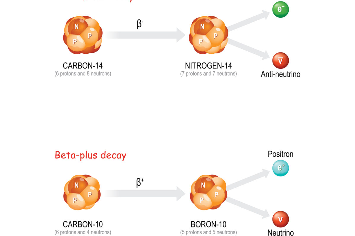
The gamma camera operates on the principles of gamma-ray detection and image formation. The process begins with the administration of a radiopharmaceutical, a compound labelled with a radioactive isotope, to the patient. This radiopharmaceutical accumulates in specific organs or tissues, emitting gamma rays as they decay.
The gamma camera’s scintillation crystal detects the emitted gamma rays, typically made of sodium iodide doped with thallium. When gamma rays interact with the crystal, they produce flashes of light or scintillations. These flashes are then converted into electrical signals by an array of photomultiplier tubes (PMTs) positioned behind the crystal.
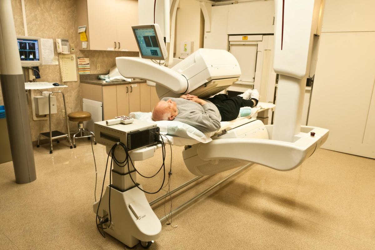
The electrical signals generated by the PMTs are processed to determine the position and intensity of the gamma ray interactions within the crystal. By compiling these data points, the gamma camera constructs a two-dimensional image that represents the spatial distribution of the radiopharmaceutical within the body. Advanced techniques, such as single-photon emission computed tomography (SPECT), allow for the generation of three-dimensional images by acquiring multiple two-dimensional images from different angles.
Clinical Applications of the Medical Gamma Camera
The gamma camera’s versatility makes it invaluable in diagnosing and managing a wide range of medical conditions. Its applications span various medical specialties, including cardiology, oncology, and neurology.
Cardiology
In cardiology, the gamma camera is instrumental in myocardial perfusion imaging. This technique assesses blood flow to the heart muscle, helping to diagnose coronary artery disease and evaluate the effectiveness of treatments such as angioplasty or bypass surgery. By imaging the distribution of radiopharmaceuticals like technetium-99m or thallium-201, clinicians can identify areas of reduced blood flow indicative of ischemia or infarction.
Oncology
Oncology is another field where the gamma camera plays a crucial role. It is used in bone scintigraphy to detect metastatic bone disease, assess the spread of cancer, and monitor treatment response. Furthermore, the gamma camera aids in localising primary tumours and evaluating lymphatic spread through sentinel lymph node imaging.
Neurology
In neurology, the gamma camera is employed in brain imaging to diagnose and manage conditions such as epilepsy, Alzheimer’s disease, and Parkinson’s disease. By visualising the distribution of radiopharmaceuticals such as fluorodeoxyglucose (FDG) or technetium-99m, clinicians can assess brain function and identify abnormalities related to these neurological disorders.
Other Applications
Beyond these specialties, the gamma camera is utilised in renal imaging to evaluate kidney function, hepatobiliary imaging to assess liver and gallbladder function, and thyroid imaging to diagnose thyroid disorders. Its ability to provide functional information complements anatomical imaging techniques, offering a more comprehensive view of the patient’s condition.
Advantages of Gamma Camera Imaging
The gamma camera offers several advantages that contribute to its widespread medical imaging use.
Non-Invasive Nature
One of the primary benefits is its non-invasive nature. Gamma camera imaging involves the administration of radiopharmaceuticals, which generally have a favourable safety profile and minimal side effects. This makes it a patient-friendly diagnostic tool suitable for repeated use in monitoring disease progression or treatment response.
Functional Imaging
Unlike other imaging modalities that primarily provide anatomical information, the gamma camera excels in functional imaging. It allows clinicians to observe physiological processes in real-time, offering insights into organ function, blood flow, and metabolic activity. This functional perspective is particularly valuable in early disease detection, where anatomical changes may not yet be apparent.
Quantitative Analysis
Gamma camera imaging enables quantitative analysis, providing measurable data on the distribution and concentration of radiopharmaceuticals within the body. This quantitative aspect enhances the accuracy of diagnoses and aids in treatment planning and monitoring. For instance, quantitative assessment of myocardial perfusion in cardiology can guide decisions regarding revascularisation procedures.
Challenges and Limitations
Despite its numerous advantages, the gamma camera has some limitations that need to be considered.
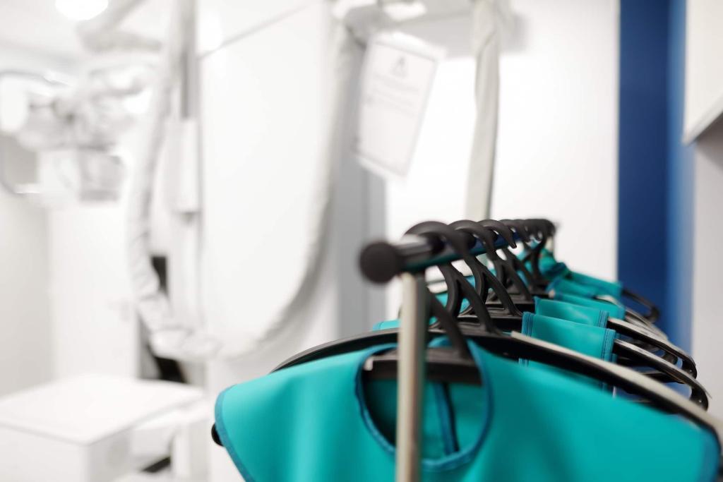
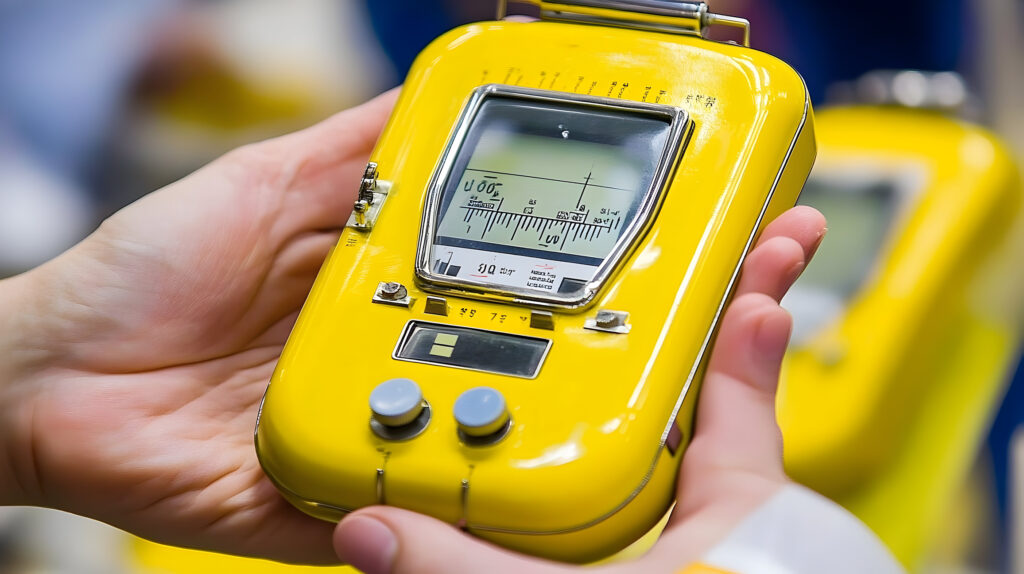
One of the primary concerns associated with gamma camera imaging is radiation exposure. Although the radiation doses used are generally low and within safe limits, cumulative exposure from multiple studies can pose risks. Therefore, careful consideration is required to balance the diagnostic benefits against the potential harm from radiation.
The resolution and sensitivity of gamma camera images are inherently lower compared to other imaging modalities, such as magnetic resonance imaging (MRI) or computed tomography (CT). This limitation can affect the detection of small lesions or subtle abnormalities. Advances in technology, such as the development of high-resolution gamma cameras and hybrid imaging systems (e.g., SPECT/CT), aim to address these challenges.
The cost and accessibility of gamma camera imaging can also be limiting factors. The equipment and radiopharmaceuticals are expensive, and the availability of gamma camera services may be limited in certain regions. Additionally, the specialised training required for personnel to operate the equipment and interpret the images can pose challenges in resource-limited settings.
Future Prospects
The future of gamma camera imaging looks promising, with ongoing advancements to improve its capabilities and expand its applications.
Technological innovations are continuously enhancing the performance of gamma cameras. The development of new scintillation materials, such as cadmium zinc telluride (CZT), offers improved energy resolution and sensitivity. This advancement allows for better image quality and more accurate quantification of radiopharmaceutical distribution. Additionally, the integration of artificial intelligence (AI) and machine learning algorithms holds potential for automated image analysis and improved diagnostic accuracy.
Hybrid Imaging Systems
Hybrid imaging systems, such as SPECT/CT and positron emission tomography (PET)/CT, combine the strengths of different imaging modalities to provide comprehensive diagnostic information. These systems allow for the simultaneous acquisition of functional and anatomical images, enhancing diagnostic accuracy and treatment planning. The fusion of gamma camera imaging with other modalities opens new avenues for research and clinical applications.
Radiopharmaceutical Development
Advances in radiopharmaceutical development are expanding the range of tracers available for gamma camera imaging. Novel radiotracers targeting specific molecular pathways and receptors offer improved specificity and sensitivity for various diseases. For example, the development of radiotracers for imaging neuroinflammation holds promise for diagnosing and monitoring neurodegenerative diseases.
Personalised Medicine
Gamma camera imaging is expected to play a significant role in personalised medicine. Providing detailed information on individual patients’ physiological processes enables tailored treatment strategies. For instance, in oncology, gamma camera imaging can help identify patients who are likely to respond to specific therapies, guiding personalised treatment plans and improving outcomes.
Conclusion
The gamma camera has become an indispensable tool in medical imaging, offering unique insights into physiological processes and aiding in diagnosing and managing a wide range of medical conditions. Its non-invasive nature, functional imaging capabilities, and quantitative analysis provide significant advantages in clinical practice. While challenges such as radiation exposure, resolution limitations, and cost persist, ongoing advancements in technology and radiopharmaceutical development are poised to address these issues and expand the gamma camera’s potential.
As the field of nuclear medicine continues to evolve, the gamma camera’s role is likely to grow, contributing to more accurate diagnoses, personalised treatment strategies, and improved patient outcomes. Embracing these advancements and addressing the associated challenges will ensure that the gamma camera remains a cornerstone of modern medical imaging.
Disclaimer
The content of this article, The Role of the Medical Gamma Camera in Modern Healthcare, published by Open Medscience on 7 July 2024, is intended for informational and educational purposes only. It is not intended to serve as medical advice, diagnosis, or treatment and should not be relied upon as a substitute for consultation with qualified medical professionals. While every effort has been made to ensure the accuracy and currency of the information provided, Open Medscience makes no guarantees regarding the completeness, reliability, or applicability of the content.
Any views expressed in the article are those of the authors and do not necessarily reflect the official policy or position of Open Medscience. Readers are encouraged to consult healthcare professionals before making any decisions based on the material presented. Open Medscience does not accept responsibility for any consequences arising from the use or misuse of the information contained herein.
home » blog » medical physics »