SPECT imaging, combined with radiotracers, delivers detailed insights into the body’s functional processes, enhancing diagnostic accuracy and personalised treatment.
SPECT-CT: A Hybrid Approach to Functional and Anatomical Imaging
- SPECT-CT hybrid scanners allow for integrating the functional information obtained from SPECT with the anatomical information obtained from CT. This results in more accurate localisation of the radiotracer and improved image quality.
- Conventional SPECT detectors use scintillation crystals to detect gamma rays, but newer SPECT systems use cadmium zinc telluride (CZT) detectors. CZT detectors have higher sensitivity and energy resolution, so they can detect and differentiate photons more accurately, resulting in better image quality.
- TOF technology improves the temporal resolution of SPECT imaging. By measuring the time taken for a gamma ray to travel from the radiotracer to the detector, TOF technology can more accurately determine the location of the radiotracer and produce better images.
- SPECT images are reconstructed from the data acquired by the detectors. Iterative reconstruction algorithms use statistical methods to iteratively refine the reconstructed image iteratively, resulting in improved image quality and reduced imaging time.
- The development of new radiotracers has expanded the clinical applications of SPECT imaging. Radiotracers are molecules labelled with a radioactive isotope and used to target specific organs or tissues. New radiotracers have been developed for various applications, such as detecting Alzheimer’s, cancer, and cardiac diseases.
These technological advances have made SPECT imaging a more accurate and efficient diagnostic tool, enabling earlier disease detection and improved treatment outcomes.
Multi-Detector SPECT Scanner Configurations
Several types of SPECT scanners can be classified based on their design, configuration, and imaging capabilities. These scanners are capable of implementing the following diagnostic imaging:
- Dedicated SPECT scanners are designed for specific imaging applications, such as cardiac or brain SPECT. They are optimised for high sensitivity and resolution in the targeted area of the body.
- SPECT scanners are designed to perform a variety of imaging studies across different body parts. They are flexible and can be used for various clinical applications.
- Stand-alone SPECT scanners comprise a gamma camera and a computer system for image reconstruction. They are typically used in smaller clinics or medical facilities.
- SPECT-CT hybrid scanners combine SPECT with CT (Computed Tomography) technology to provide functional and anatomical imaging in a single scan. They offer improved accuracy and specificity by combining the benefits of both technologies.
- SPECT-MRI hybrid scanners combine SPECT with MRI (Magnetic Resonance Imaging) technology to provide functional and anatomical imaging in a single scan. They are particularly useful for studying brain disorders and other conditions that require high spatial resolution.
- Mobile SPECT scanners are designed to be transported to various locations for on-site imaging studies. They are often used in emergency departments, intensive care units, and other clinical settings requiring rapid imaging.
Each SPECT scanner type has unique advantages and disadvantages, depending on the clinical application and imaging requirements.
CZT SPECT Scanners: Improving Image Quality and Reducing Radiation Dose
The most advanced is the CZT-based SPECT scanner. CZT stands for cadmium zinc telluride, a semiconductor material used as the detector material in these scanners. CZT-based SPECT scanners have several advantages over conventional SPECT scanners.
CZT detectors are more sensitive to gamma rays than conventional scintillation detectors. This means that CZT-based SPECT scanners require less radiation dose to obtain high-quality images. These detectors have better energy resolution than conventional scintillation detectors and allow CZT-based SPECT scanners to distinguish between photons of different energy levels, resulting in higher image quality and better diagnostic accuracy.
Also, CZT-based SPECT scanners can acquire images faster than conventional SPECT scanners. This means that patients spend less time undergoing the scan, and the imaging department can serve more patients in a given time period. In addition, CZT detectors have better spatial resolution than conventional scintillation detectors and can produce more detailed images, which is particularly useful for small lesions or structures.
One example of a CZT-based SPECT scanner is the Discovery NM/CT 670 CZT scanner developed by GE Healthcare. This scanner uses CZT detectors and advanced reconstruction algorithms to produce high-quality images with improved spatial resolution and diagnostic accuracy.
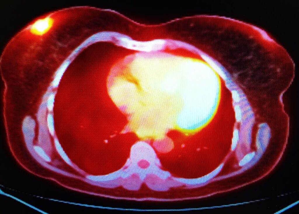
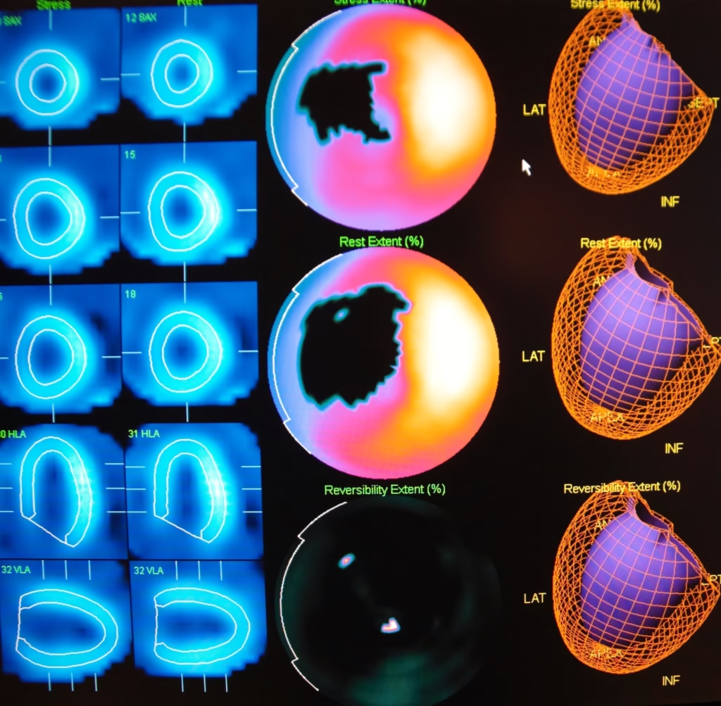
SPECT scanners are used for various medical imaging applications and use radiotracers, which are molecules labelled with a radioactive isotope and injected into the patient’s bloodstream. The radiotracer emits gamma rays detected by the SPECT scanner, allowing for the production of 3-D images of the inner structures and functions of the body. The areas of clinical applications of SPECT imaging include studying various brain disorders, such as Alzheimer’s disease, Parkinson’s disease, and epilepsy. It can help diagnose, stage, and monitor these conditions. Also used to evaluate blood flow in the heart and diagnose heart conditions, such as coronary artery disease and heart failure. It may also be used to assess the effectiveness of treatment and predict the risk of future heart problems.
Furthermore, SPECT imaging is used to detect and stage cancer and monitor the response to treatment, including localising tumours and guiding biopsy procedures. Moreover, SPECT imaging can evaluate bone disorders such as fractures, infections, and tumours.
Radiopharmaceuticals in SPECT Imaging
Radiopharmaceuticals contain a radioactive tracer and are used in medical imaging to diagnose and treat diseases. For example, in SPECT imaging, these radiotracers emit gamma rays and are detected by the SPECT scanner to create 3-D images of internal organs and tissues. The following radiotracers are used in SPECT imaging:
- Technetium-99m (Tc-99m, t1/2= 6 hrs) is the most commonly used radiopharmaceutical in SPECT imaging. It is attached to various compounds depending on the organ or tissue being imaged, such as the heart, brain, bone, and kidneys.
- Iodine-123 (I-123, t1/2= 13.2 hrs) is used to image the thyroid gland and often diagnoses thyroid disorders such as hyperthyroidism and hypothyroidism.
- Thallium-201 (Tl-201, t1/2= 73 hrs ) is used to evaluate blood flow to the heart muscle and can help diagnose coronary artery disease.
- Gallium-67 (Ga-67, t1/2= 78 hrs) is used to detect inflammation and infection in the body and is often used in diagnosing and monitoring cancer.
- Indium-111 (In-111, t1/2= 67 hrs) is used to evaluate inflammation and infection in the body and can also image certain types of tumours.
Radiotracers used in SPECT imaging are generally safe and well tolerated, but as with any medical procedure involving radiation, there is a small risk of radiation exposure. Therefore, the scanning procedures are discussed with the patient before imaging is performed.
Next-Generation SPECT Systems: Enhancing Sensitivity, Resolution, and Speed
The future of SPECT imaging will likely involve advancements in hardware and software that will improve image quality and increase the accuracy and specificity of SPECT scans.
One area of development in SPECT imaging is new radiotracers, which are substances injected into the body and emit gamma rays that the SPECT scanner can detect. Researchers are exploring the use of radiotracers specific to certain types of cells or tissues, which could improve the ability of SPECT scans to detect and diagnose diseases.
Another development area is using hybrid imaging techniques that combine SPECT with other imaging modalities, such as computerised Tomography (CT) or magnetic resonance imaging (MRI). These hybrid techniques can provide more detailed and precise images by combining the strengths of each imaging modality.
Advancements in SPECT hardware and software are also likely to play a role in the future of SPECT imaging. For example, improvements in detector technology could increase the sensitivity and resolution of SPECT scanners, while advancements in image reconstruction algorithms could improve image quality and reduce image artefacts.
Disclaimer
The content provided in “SPECT Imaging Technology: From Single Photon Emission Tomography to Hybrids” is intended for informational and educational purposes only. It does not constitute medical advice, diagnosis, or treatment, and should not be relied upon as a substitute for consultation with qualified healthcare professionals.
While every effort has been made to ensure the accuracy of the information presented, Open Medscience makes no warranties or representations regarding its completeness, reliability, or applicability in clinical practice. The technologies, scanners, radiopharmaceuticals, and procedures described are subject to ongoing research and regulatory review and may not be available or suitable for all clinical settings or patients.
Readers are strongly encouraged to consult qualified professionals for guidance regarding the use of SPECT imaging or any other medical technologies discussed. Open Medscience accepts no liability for any decisions made based on this content or for any actions taken or not taken as a result of reading this article.
Use of this information is at your own risk.
home » blog » nuclear medicine imaging »