Positron Emission Tomography (PET) scans are sophisticated imaging tools used in medical diagnostics to observe metabolic processes in the body. This brief guide, “How to Read a PET Scan”, looks into the principles behind PET scans, the preparation required, the scanning process, and the interpretation of results. By understanding these aspects, patients and medical professionals can gain insights into how PET scans aid in diagnosing and managing various conditions, including cancer, neurological disorders, and cardiovascular diseases.
Introduction to PET Scans
Positron Emission Tomography (PET) scans are advanced imaging techniques that provide detailed pictures of the metabolic activity within the body. Unlike other imaging modalities that primarily depict anatomical structures, PET scans offer functional insights, making them invaluable in diagnosing and monitoring various medical conditions.
PET scans involve the use of a radiotracer, a substance that emits positrons as it decays. The most commonly used radiotracer is fluorodeoxyglucose (FDG), a glucose analogue. When injected into the body, FDG accumulates in areas with high metabolic activity, such as cancer cells. As the FDG decays, it emits positrons that collide with electrons, producing gamma rays. The PET scanner detects these gamma rays to create detailed images of metabolic processes.
A PET scanner consists of a ring of detectors that capture the gamma rays emitted from the body. A computer then processes the data collected to generate cross-sectional images, often combined with CT or MRI scans for enhanced anatomical reference.
Preparing for a PET Scan
To ensure accurate results, patients are given specific instructions to follow before a PET scan. These may include fasting for several hours, avoiding strenuous physical activity, and disclosing any medications or medical conditions. Adhering to these guidelines helps optimise the quality of the images.
The radiotracer is injected into the patient’s bloodstream on the day of the scan. The patient then waits for about 30 to 60 minutes to allow the tracer to distribute throughout the body. During this period, it is crucial to remain still and relaxed to prevent unnecessary movement, which can affect the scan’s accuracy.
During the Scan
The patient lies on a motorised table that slides into the PET scanner’s gantry. The scan itself typically lasts between 20 to 40 minutes, during which the patient must remain as still as possible to ensure clear images. The scanner may produce clicking or buzzing noises, but the procedure is painless.
Post-Scan Care
After the scan, patients are advised to drink plenty of fluids to help flush the radiotracer from their system. Normal activities can usually be resumed immediately, although specific instructions may be given based on the patient’s condition and the purpose of the scan.
How to Read a PET Scan
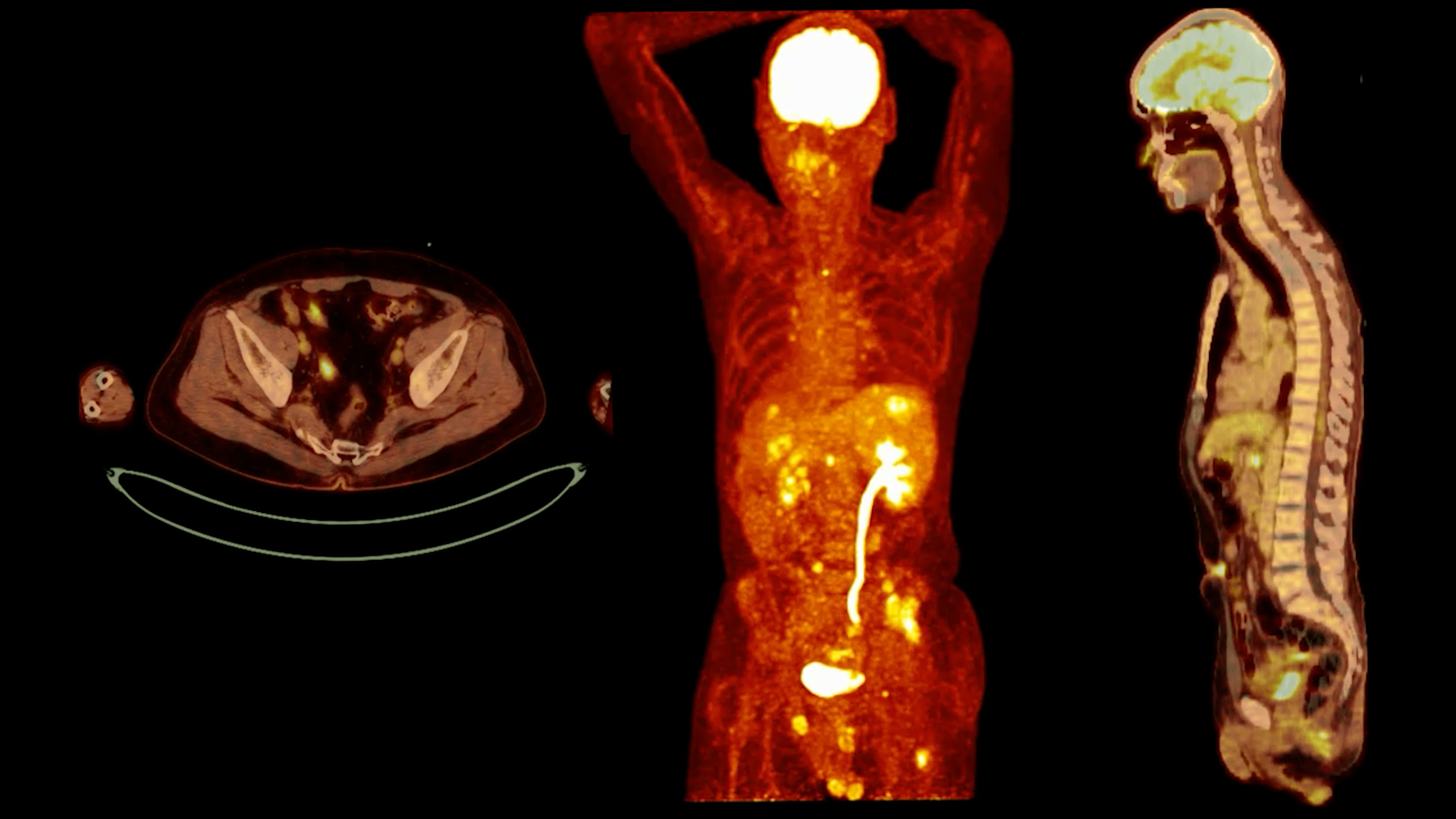
Radiologists or nuclear medicine specialists interpret PET scan images. The images display varying levels of radiotracer uptake, indicating different metabolic activities. Areas with high uptake appear brighter, while areas with low uptake appear darker.
Key Indicators
- Hypermetabolic Activity: High radiotracer uptake suggests increased metabolic activity, often associated with tumours, inflammation, or infection.
- Hypometabolic Activity: Low radiotracer uptake indicates decreased metabolic activity, which can be seen in conditions like Alzheimer’s disease or areas of tissue damage.
Table of PET Scan Colours and Their Meanings
| Colour | Metabolic Activity | Interpretation |
| Red | Very High | High radiotracer uptake often indicates active metabolic processes such as cancer cells or inflammation. |
| Orange | High | Elevated metabolic activity can suggest areas of infection, inflammation, or tumours. |
| Yellow | Moderate to High | Intermediate level of radiotracer uptake, potentially indicating regions of concern that require further investigation. |
| Green | Moderate | Normal metabolic activity generally indicates healthy tissue or a stable condition. |
| Blue | Low | Normal metabolic activity, generally indicates healthy tissue or a stable condition. |
| Black | Very Low or None | Minimal to no radiotracer uptake, often indicating areas of necrosis, infarction, or tissues that are not metabolically active. |
Applications of PET Scans
Oncology
PET scans are widely used in oncology for detecting and staging cancers, evaluating treatment response, and monitoring for recurrence. They can identify malignant cells that may not be visible on other imaging tests, aiding in early diagnosis and precise treatment planning.
Neurology
In neurology, PET scans help diagnose and manage neurological disorders such as Alzheimer’s disease, epilepsy, and Parkinson’s disease. PET scans provide valuable information about the function of different brain regions by assessing brain metabolism.
Cardiology
PET scans are also utilised in cardiology to assess myocardial viability and blood flow. They can detect areas of the heart damaged by a heart attack or at risk of future cardiovascular events, guiding treatment decisions.
Advantages and Limitations
Benefits of PET Scans
- Functional Imaging: PET scans provide unique insights into the body’s metabolic processes, complementing anatomical imaging techniques.
- Early Detection: By identifying changes at the cellular level, PET scans can detect diseases in their early stages.
- Comprehensive Evaluation: PET scans can evaluate the entire body in a single session, offering a holistic view of a patient’s condition.
Limitations of PET Scans
- Radiation Exposure: The use of radiotracers involves exposure to radiation, although the levels are generally low and considered safe.
- Availability and Cost: PET scans are complex and expensive, limiting their availability in some regions.
- False Positives/Negatives: PET scans can sometimes produce false results, requiring further confirmation through other diagnostic tests.
Enhancing PET Scan Accuracy
PET scans are often combined with CT or MRI scans to improve diagnostic accuracy. Functional and anatomical imaging fusion allows for precise localisation and characterisation of abnormalities.
Continuous advancements in PET scan technology, such as the development of new radiotracers and improved scanner resolution, are enhancing the accuracy and utility of PET scans. These innovations are expanding the range of conditions that can be diagnosed and monitored effectively.
Practical Considerations for Patients
Patients should openly communicate with their healthcare providers about any concerns or questions regarding the PET scan procedure. Understanding the purpose of the scan, what to expect, and how to prepare can alleviate anxiety and ensure a smoother experience.
After the scan, patients should discuss the results with their healthcare provider. The interpretation of PET scan images requires specialised knowledge, and a detailed explanation can help patients understand their diagnosis and treatment options.
Future Directions of PET Scanning
Ongoing research is focused on developing new radiotracers targeting specific diseases and conditions. These advancements aim to improve the specificity and sensitivity of PET scans, enabling earlier and more accurate diagnosis.
As our understanding of metabolic processes deepens, the clinical applications of PET scans are expanding. Emerging uses include monitoring the effectiveness of novel therapies, guiding surgical planning, and assessing the progression of chronic diseases.
Conclusion
PET scans are highly effective diagnostic tools that offer detailed insights into the body’s metabolic processes. By understanding how these scans operate, how to prepare for them, and how to interpret the results, patients and healthcare professionals can make the most of their capabilities. Although they have certain limitations, their role in early detection, accurate diagnosis, and thorough assessment makes them an essential part of modern medical practice. Continuing research and technological progress are set to improve their performance even further.
Disclaimer
The content provided in this article, How to Read a PET Scan: A Basic Understanding, is intended for general informational purposes only and should not be considered medical advice, diagnosis, or a substitute for consultation with a qualified healthcare professional. Open Medscience makes every effort to ensure the accuracy of the information presented; however, no guarantee is made regarding its completeness, relevance, or timeliness.
PET scan interpretation requires specialised training and should always be performed by appropriately qualified radiologists or nuclear medicine specialists. Patients should not attempt to interpret their own scan results and are strongly advised to discuss all medical concerns, diagnoses, and treatment options with their doctor or medical team.
Open Medscience accepts no responsibility for any loss, injury, or damage resulting from the use of the information contained in this article. Use of this material is at the reader’s own discretion and risk.
home » blog » features »