Echocardiogram
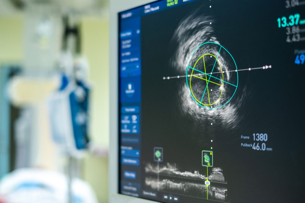
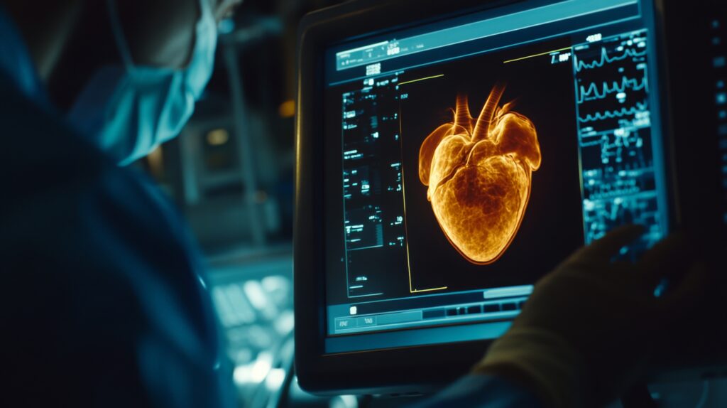
An echocardiogram is a non-invasive medical test that uses high-frequency sound waves to produce detailed images of the heart. It is also known as an echo test or cardiac ultrasound. A transducer is placed on the patient’s chest during an echocardiogram. The transducer emits sound waves that bounce off the heart and create echoes. These echoes are then recorded by the transducer and processed by a computer to create images of the heart’s structure and function.
Echocardiography is a versatile diagnostic tool that can detect various heart conditions, such as heart valve disease, heart failure, and congenital heart defects. It can be used to evaluate the heart’s pumping function, measure the size of the heart’s chambers, and assess blood flow through the heart. There are several types of echocardiograms, each with its specific purpose:
Transthoracic echocardiogram (TTE): This is the most common type of echocardiogram. It involves placing the transducer on the patient’s chest and producing images of the heart from outside the body. Transesophageal echocardiogram (TEE) involves inserting a thin, flexible tube with a transducer attached down the patient’s throat and into the oesophagus. This allows for more detailed images of the heart, as the oesophagus lies directly behind the heart.
Stress echocardiogram: This involves performing an echocardiogram before and after the patient exercises or is given medication to simulate exercise. This can help diagnose heart problems that may only occur during physical activity. 3D echocardiogram: This uses specialized software to create a three-dimensional heart image. This can provide even more detailed information about the heart’s structure and function.
Echocardiograms are generally safe and non-invasive, with no known risks or side effects. In addition, they are painless and typically take between 30 and 60 minutes to complete. Therefore, echocardiography is a valuable diagnostic tool that allows doctors to visualize the heart’s structure and function in detail. It can diagnose various heart conditions and is generally safe and non-invasive. If you are experiencing symptoms such as chest pain or shortness of breath, your doctor may recommend an echocardiogram to evaluate your heart health.
You are here:
home » echocardiogram


