PET Imaging for Treatment Guidance
Positron Emission Tomography (PET) imaging has revolutionised medical diagnostics and treatment guidance by providing detailed insights into physiological processes at the molecular level. This advanced imaging technique plays a critical role in modern medicine, particularly in oncology, neurology, and cardiology, offering unparalleled precision in evaluating diseases and guiding therapeutic decisions.
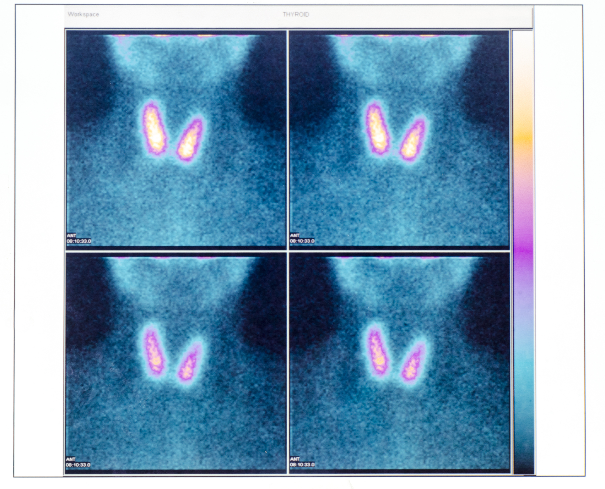
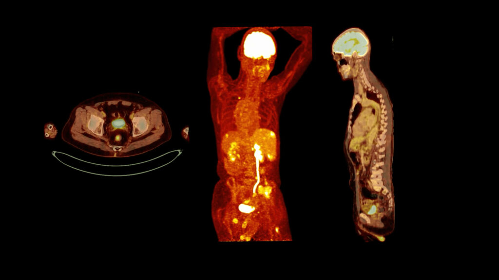
A PET scan involves the use of radiotracers, which are compounds labelled with positron-emitting radionuclides. These radiotracers are typically designed to target specific biological pathways or molecules. For example, fluorodeoxyglucose (FDG) is a commonly used radiotracer that mimics glucose, enabling the visualisation of glucose metabolism in tissues. Once administered to the patient, the radiotracer accumulates in areas of high metabolic activity, such as tumours, which are then detected by the PET scanner. The scanner measures the emitted gamma rays resulting from positron-electron annihilation, producing a three-dimensional image of the distribution of the radiotracer.
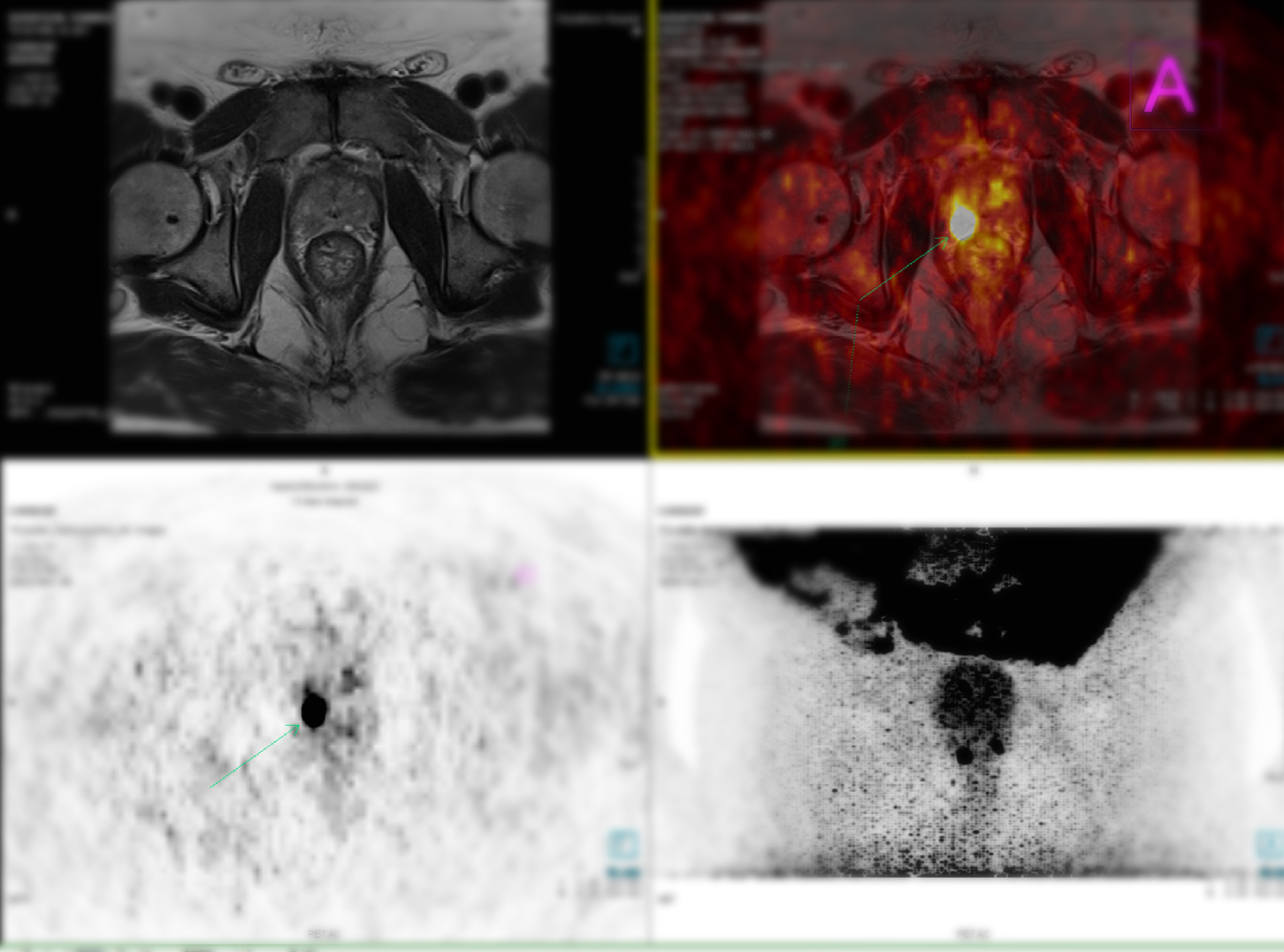
In oncology, PET imaging is invaluable for diagnosis, staging, and treatment planning. By identifying areas of abnormal metabolic activity, it aids in differentiating between benign and malignant lesions. This is particularly useful in cancers such as lymphoma, lung, and colorectal cancers. Moreover, PET scans help assess the extent of disease spread (metastasis), which is crucial for selecting appropriate treatment modalities. For example, a PET scan can identify patients who are suitable for surgery versus those who require systemic therapies such as chemotherapy or immunotherapy.
During treatment, PET imaging is employed to monitor therapeutic response, enabling clinicians to make real-time adjustments to the treatment plan. A decline in the uptake of FDG, for instance, indicates reduced metabolic activity in tumours, suggesting that the therapy is effective. Conversely, persistent high uptake may signal resistance to treatment, prompting a change in strategy. This capability to assess treatment efficacy early minimises unnecessary exposure to ineffective therapies, improving patient outcomes and reducing healthcare costs.
In neurology, PET imaging assists in diagnosing and managing conditions like Alzheimer’s disease, epilepsy, and Parkinson’s disease. Mapping brain function and identifying areas of abnormal activity support the precise localisation of pathology. Similarly, in cardiology, PET imaging evaluates myocardial viability and perfusion, aiding decisions about interventions such as revascularisation.
The future of PET imaging lies in the development of novel radiotracers targeting a wider range of diseases and the integration of PET with other imaging modalities such as CT and MRI. These advancements promise even greater accuracy in treatment guidance, personalised medicine, and improved patient care.
In conclusion, PET imaging is a cornerstone of modern medicine, providing critical insights that empower clinicians to make informed decisions. Its role in treatment guidance ensures that patients receive the most effective and tailored therapies, underscoring its significance in advancing healthcare.
home » PET Imaging for Treatment Guidance
 By
By