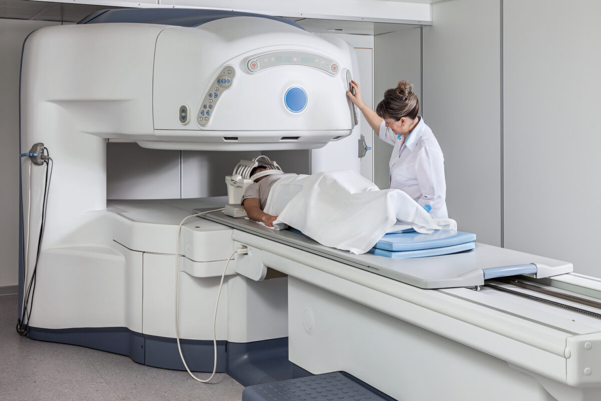
From Bedside to Ambulance: The New Era of Mobile CT Brain Imaging
 By
By
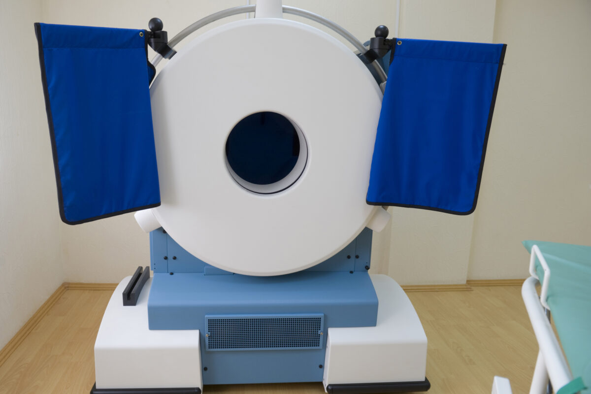
Mobile CT brain imaging is revolutionizing stroke care. Discover how this technology is used in various medical settings.
By
Mobile CT brain imaging is revolutionizing stroke care. Discover how this technology is used in various medical settings.
 By
By
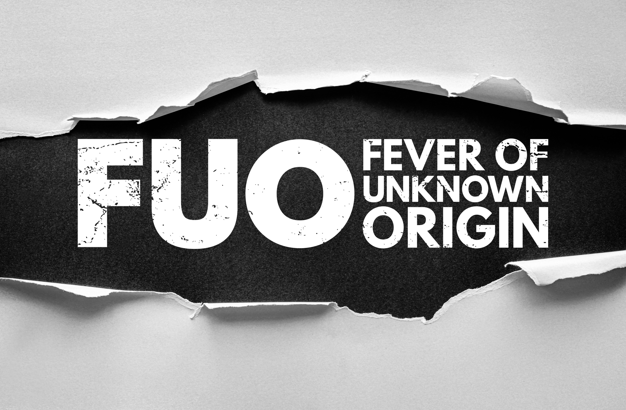
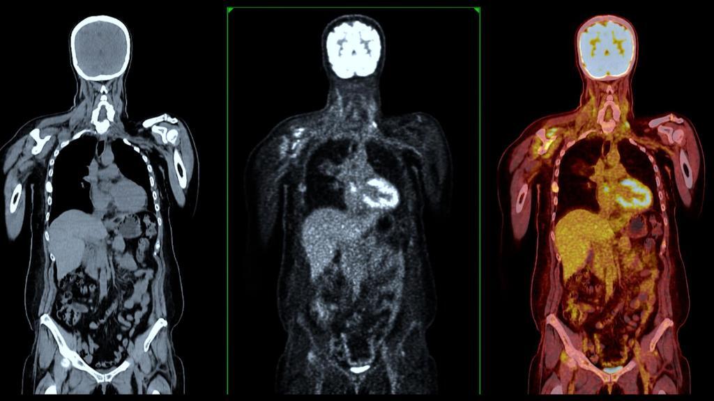
Uncover the benefits of using FDG PET-CT in fever of unknown origin and its impact on diagnostic accuracy and patient care.
 By
By
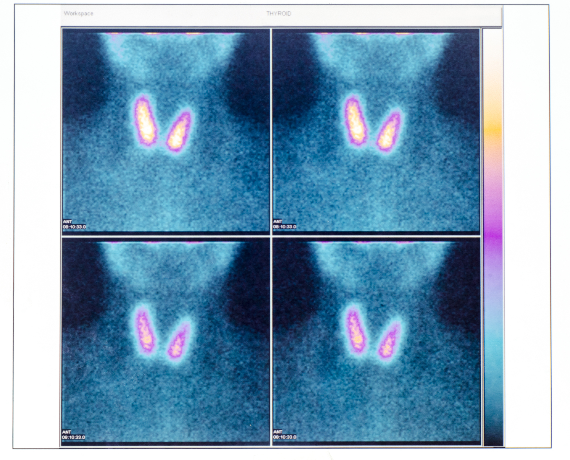
Uncover how PET/CT enhances thyroid cancer diagnosis. Explore its insights into tumours, staging, and personalised oncology.
 By
By
Discover the role of generative artificial intelligence in medical imaging, improving workflows and data quality in healthcare.
 By
By
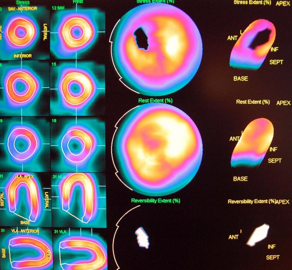
Cardiac imaging enables precise assessment of heart structure, function, and pathology using advanced non-invasive diagnostic techniques.
 By
By
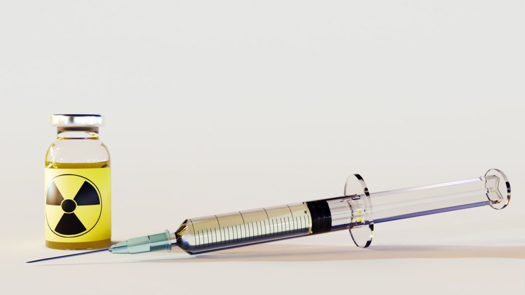
PET imaging agents play a critical role in modern medicine, enabling precise visualisation and quantification of biological processes for accurate disease diagnosis.
 By
By
Medical imaging crucially enhances oncology, aiding early cancer detection and effective treatment planning. Image for illustration only. People depicted are models.
 By
By
Pancreatic cancer demands accurate prognosis, improved treatment strategies, and PET radiomic analysis for optimised post-resection outcomes.
 By
By
Fluorine-18 Sodium Fluoride (18F-NaF) is an effective, safe diagnostic radiopharmaceutical, revolutionising bone imaging and detecting skeletal abnormalities with high accuracy.
By
Fluorine-18 Piflufolastat, a novel radiotracer, enables precise imaging of prostate cancer, enhancing diagnostic accuracy and facilitating personalied, targeted treatment approaches.
 By
By
A case report to analyse the role of abiraterone and volumetric-modulated arc therapy towards prostate cancer.