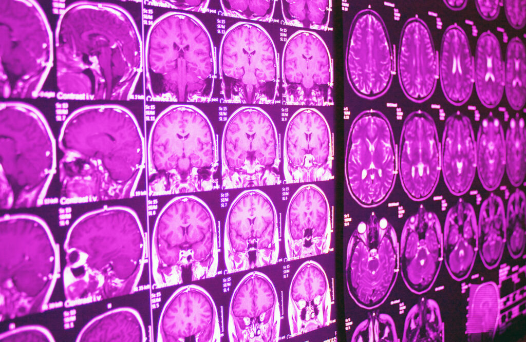
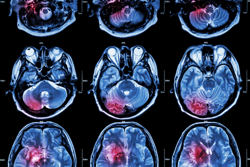
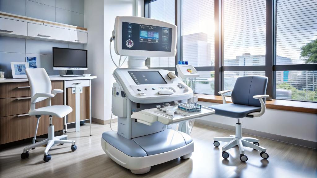
Magnetic Resonance Imaging (MRI) is one of the most advanced imaging techniques in medical diagnostics. This non-invasive technology uses magnetic fields and radio waves to produce detailed images of the body’s internal structures, offering unparalleled clarity for soft tissues, organs, and bones. This article provides a brief overview of the basics of MRI, including its working principles, components, types, applications, safety considerations, and future advancements.
Introduction to the Basics of MRI
Magnetic Resonance Imaging (MRI) is a cornerstone of modern medical diagnostics, providing detailed, high-resolution images of the human body’s internal structures. Unlike X-rays or CT scans, MRI does not use ionising radiation, making it a safer choice for repeated imaging. Since its inception in the early 1970s, MRI has transformed how clinicians diagnose and monitor various conditions, from neurological disorders to musculoskeletal injuries.
The Science Behind MRI
The basics of MRI are based on the principles of nuclear magnetic resonance (NMR), a phenomenon discovered in the 1940s. It exploits the magnetic properties of atomic nuclei, primarily hydrogen, which are abundant in water and fat within the human body. When placed in a strong magnetic field, these nuclei align with the field and can absorb energy from specific radiofrequency pulses. Upon returning to their equilibrium state, they emit energy detectable by MRI scanners.
Key Concepts in MRI Physics
- Protons and Magnetic Fields: Hydrogen nuclei contain a single proton that acts like a tiny magnet. In the presence of a magnetic field, these protons align with or against the field’s direction.
- Resonance: When radiofrequency (RF) energy matches the protons’ natural frequency, they absorb the energy, a process called resonance.
- Relaxation: After the RF pulse stops, the protons return to their original alignment, releasing energy in two forms:
- T1 Relaxation (Longitudinal): The time it takes for protons to realign with the magnetic field.
- T2 Relaxation (Transverse): The time it takes for protons to lose synchronisation with each other.
Components of an MRI Scanner
Magnet
The magnet is the core of an MRI scanner and generates the powerful magnetic field required for imaging. Most MRI machines use superconducting magnets cooled with liquid helium to maintain high magnetic strength, typically 1.5 to 3 Tesla (T).
Gradient Coils
Gradient coils are secondary magnets that create spatial variations in the magnetic field, enabling the localisation of signals. These gradients are crucial for constructing images slice by slice.
Radiofrequency Coils
RF coils act as both transmitters and receivers. They send RF pulses to excite protons and detect the emitted signals.
Computer System
A sophisticated computer processes the detected signals into detailed images, using algorithms to reconstruct data into cross-sectional or 3D representations.
Types of MRI
Structural MRI
This type provides static images of anatomical structures and is used to detect abnormalities like tumours, fractures, and organ damage. Common sequences include:
- T1-Weighted Images: Excellent for anatomical details.
- T2-Weighted Images: Ideal for detecting fluid accumulations and inflammation.
Functional MRI (fMRI)
Functional MRI measures brain activity by detecting changes in blood flow. It is widely used in neuroscience research, including studies on cognition, behaviour, and neurological disorders.
Diffusion MRI
This technique tracks the movement of water molecules within tissues, making it invaluable for identifying strokes and mapping brain white matter tracts.
Magnetic Resonance Angiography (MRA)
MRA visualises blood vessels without the need for contrast agents in some cases. It is used for diagnosing vascular diseases and aneurysms.
Cardiac MRI
Specialised protocols capture images of the heart, providing information about structure, function, and blood flow.
Applications of MRI
Neurological Imaging
MRI is the gold standard for imaging the brain and spinal cord, aiding in the diagnosis of conditions such as:
- Stroke
- Multiple sclerosis
- Brain tumours
- Degenerative disorders like Alzheimer’s disease
Musculoskeletal Imaging
MRI excels at imaging soft tissues, making it the preferred method for diagnosing:
- Ligament and tendon injuries
- Spinal disc herniations
- Bone marrow diseases
Oncological Imaging
MRI helps detect and stage cancers, particularly in soft tissues, such as the liver, prostate, and breast.
Abdominal and Pelvic Imaging
MRI provides clear images of organs like the liver, pancreas, and uterus, often used for evaluating tumours or inflammatory conditions.
Advantages of MRI
- Non-Invasive and Safe: No ionising radiation is involved.
- High Contrast Resolution: Excellent for soft tissue imaging.
- Versatility: Applicable to nearly every body part.
- Multiplanar Imaging: Ability to acquire images in multiple planes without repositioning the patient.
Challenges and Limitations
Cost and Accessibility
MRI scanners are expensive to install and maintain, limiting their availability in some regions.
Scan Duration
MRI scans are time-intensive, often requiring 30 minutes to an hour, which can be challenging for patients with claustrophobia or those who cannot remain still.
Contraindications
Certain conditions, such as the presence of metallic implants or pacemakers, may preclude MRI use.
Safety Considerations
Magnetic Field Interactions
The strong magnetic fields can attract ferromagnetic objects, posing risks to both patients and equipment. Safety protocols ensure no metallic objects enter the scanner room.
Radiofrequency Exposure
While RF energy is non-ionising, excessive exposure can cause tissue heating. Scanners have safety checks to prevent this.
Contrast Agents
Gadolinium-based contrast agents are generally safe but may pose risks in patients with severe kidney impairment.
Preparing for an MRI Scan
Pre-Scan Guidelines
Patients are typically advised to remove all metal objects and inform staff of any implants or medical devices. Loose-fitting clothing without metal fasteners is recommended.
During the Scan
The patient lies on a table that slides into the MRI scanner. Ear protection is often provided to reduce discomfort from the machine’s loud noises.
Future Directions in MRI Technology
Higher Field Strengths
MRI systems with stronger magnets (7T and beyond) promise even greater resolution and faster imaging.
Artificial Intelligence (AI) Integration
AI is being used to enhance image reconstruction, reduce scan times, and improve diagnostic accuracy.
Portable MRI Systems
Advancements in technology are enabling the development of lightweight, portable MRI scanners, increasing accessibility in remote areas.
Improved Contrast Agents
Research into non-gadolinium-based agents aims to enhance safety while maintaining image quality.
Conclusion
The basics of MRI have revolutionised medical imaging, offering unmatched clarity and safety for diagnosing a wide range of conditions. Its versatility and precision make it indispensable in modern medicine. As technology continues to evolve, MRI will likely become even more powerful and accessible, further advancing healthcare and research. Understanding its basics helps demystify this remarkable tool and highlights its vital role in patient care.
Disclaimer
The information provided in this article is for general educational purposes only and is not intended as medical advice, diagnosis, or treatment. While efforts have been made to ensure the accuracy of the content, Open Medscience does not guarantee the completeness or reliability of the information presented. Readers should consult a qualified healthcare professional for advice on any medical condition or imaging procedure. This article does not replace the guidance of medical experts or regulatory authorities. Open Medscience disclaims all liability for any loss or damage arising from reliance on the material contained herein.
home » blog » medical imaging modalities »


