The new technologies emerging in the clinical setting include fractional flow reserve (FFR)-CT, CT perfusion imaging and coronary plaque assessment.
The Role of Fractional Flow Reserve-Computed Tomography (FFR-CT)
Since introducing the standard 64-slice CT scanners, today’s cardiac computed tomography imaging can examine a patient using a very low radiation dose to obtain a cardiac scan not exceeding 3 mSv. This is only possible through the technological advancements of more sensitive detectors and faster gantry speed rotation. Also, image reconstruction software enables low-radiation dose scans to produce in-depth resolution diagnostic quality images compared to high x-ray doses in older cardiac CT scanners.
The new technologies emerging in the clinical setting include fractional flow reserve (FFR)-CT, CT perfusion imaging and coronary plaque assessment. These breakthroughs will bring CT beyond the anatomical evaluation to allow physiological evaluation without the patient undergoing nuclear perfusion scans or invasive diagnostic angiography. Especially the more advanced automated visualisation software used in an invasive examination in the cath lab where an FFR is performed and intravascular ultrasound imaging is used to evaluate the lesions. However, CT offers a non-invasive way to achieve these same assessments.
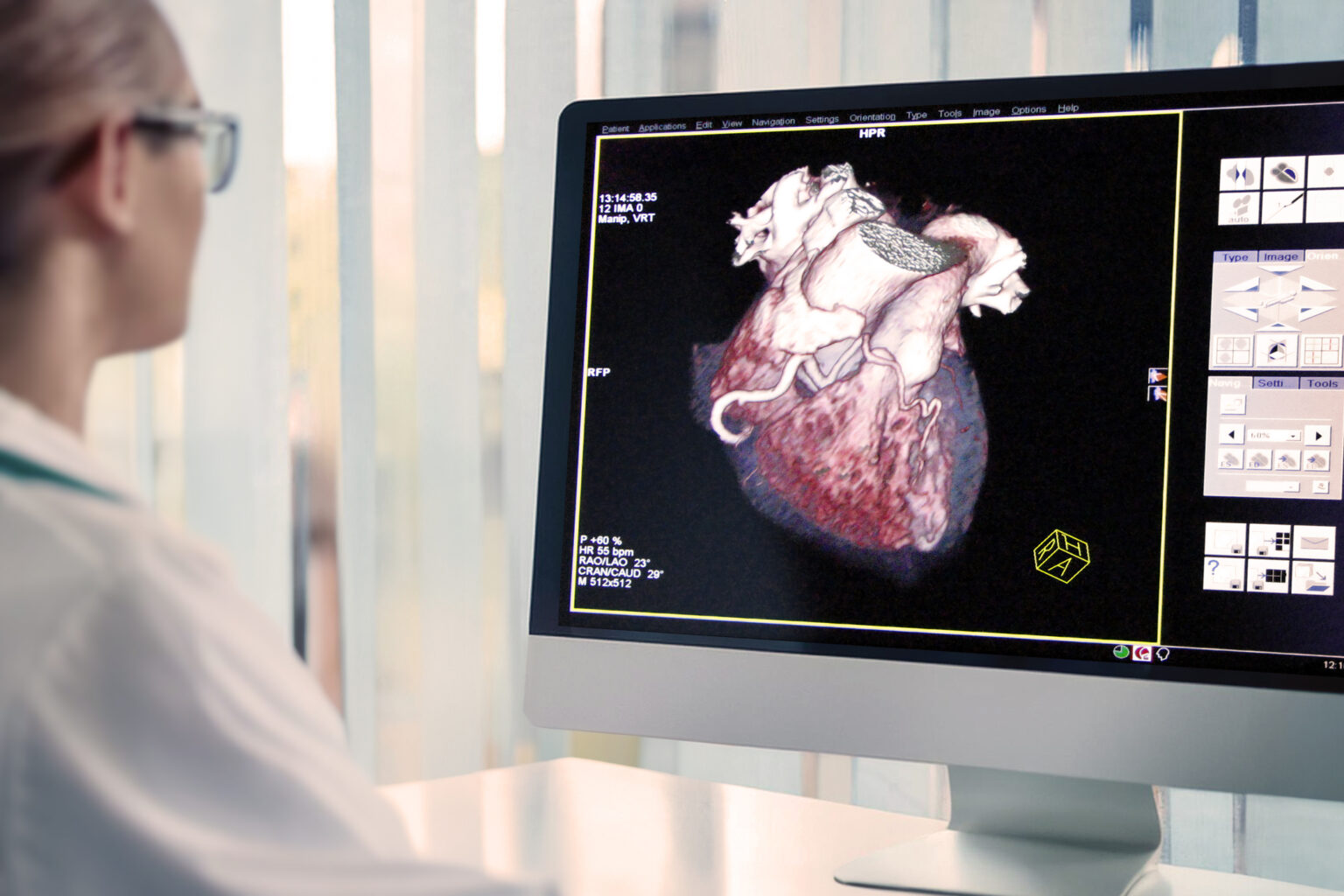
FFR-CT has been approved by the US Food and Drug Administration (FDA) for the non-invasive hemodynamic assessment of the entire coronary tree. This imaging modality can create a 3-D model of the coronary arteries using colour codes based on computational fluid dynamics. These colour codes will show low-flow areas correlated to the coronary lesions. The aim is to identify lesions that may lead to ischemic chest pain or a heart attack. However, the FFR-CT technology can take several hours to return a result. The processing time will be reduced, and FFR-CT will be broadly used in the clinical setting.
The Role of Computed Tomography in Preventative Medicine and Risk Assessment
Modern CT machines have the potential for serial imaging due to very low-dose radiation not exceeding one mSv during patient scanning. Therefore, the older 64-slice CT systems must be upgraded with more sensitive detectors, including advanced image reconstruction software.
CTCA will be the leading imaging modality to identify early coronary disease
These new coronary-CT scanners aim to identify early plaque before it leads to advanced coronary disease. Stress testing does not show early plaque until you have a reasonable amount of coronary disease compared to CT scanning. CT scanning will allow the patient to personalise the treatment plan to prevent a heart attack. Furthermore, cardiac CT as a risk assessment tool already sees growth with calcium scoring. Numerous clinical trials have shown a correlation between the amount of calcified plaque in coronary vessels and a patient’s risk for a heart attack. CT calcium scoring can also be used to convince patients to go on statin therapy and calcium scoring to be performed at least every five years.
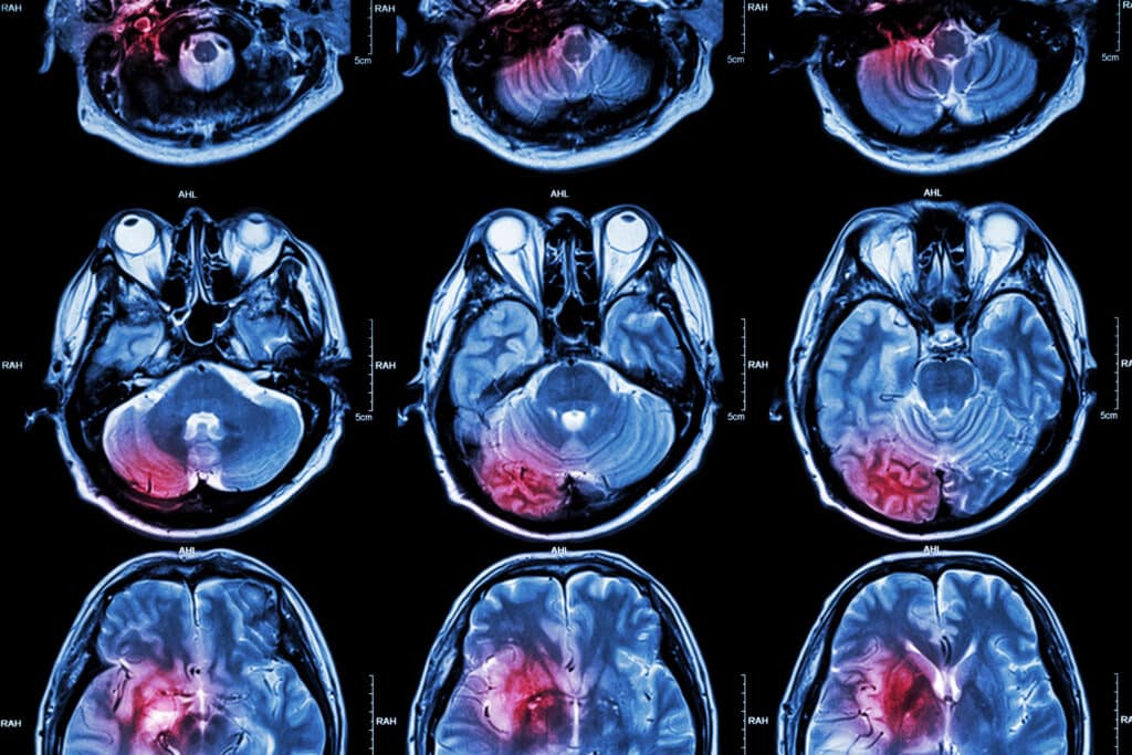
Cardiac CT Evolves: Uncovering New Areas of Usage in Cardiology
The latest CT cardiac scanners can determine structural heart evaluation and procedural planning. In addition, these scanners are also able to do precise imaging based on advanced visualisation software. These new technologies will bring state-of-the-art cardiac imaging in transcatheter aortic valve replacement (TAVR). In addition to left atrial appendage (LAA) occlusion and transcatheter mitral repair.
CT Scanners Reimagined: The Impact of Modern Technology on Medical Imaging
Over the past decade, the most significant advances in CT scanners have been delivering low-dose radiation to the patient. This can be achieved by upgrading the older CT scanners to high-slice systems, including faster gantry speeds to freeze cardiac motion. Furthermore, modern CT scanners can perform complete anatomical coverage in one rotation. The problem with the 64-slice scanners was that it usually took two rotations to image the heart. New scanner technology will lower CT cardiac exams to 3 mSv or below. However, some centres already perform routine cardiac CT exams one mSv or below using the newest scanners and image reconstruction software.
Also, the advancements in the detector resolution will increase the image sharpness and resolution by 0.2 mm. This will enable defining the smaller structures of the plaque composition and stents inside coronary vessels. In addition, the volume spectral CT technology improves tissue characterisation, small lesion detection and metal artefact reduction.
Another improvement to CT scanners is the Whisper Drive technology, allowing high-speed scans to produce diagnostic imaging of the heart in one heartbeat. This is possible using a high-speed x-ray tube that circles the patient up to five times per second. Also, a 30-degree bidirectional gantry tilt capability enables angled scanning to avoid dosing radiosensitive organs.
Furthermore, the 80-detector row (160-slice) system is designed to perform whole-body imaging and volumetric scanning. These improvements are found in the high-end CT scanners for a superior patient experience. They can provide faster reconstruction speeds of up to 50 images per second at full resolution. In addition, it optimises workflow and patient comfort with thin slices at 0.5 mm and a 78 cm bore. Moreover, these CT scanners use SURESubtraction and Single Energy Metal Artifact Reduction (SEMAR) and can be installed on 40 to 80 to 160 slices. These technological advances in CT machines enable faster scanning speeds that allow to perform of sedation-free pediatric exams and freeze cardiac motion.
Next-Generation Cardiac Imaging: The Emergence of PET and CTCA in Cardiovascular Disease Detection
Positron emission tomography (PET) scanning combined with Computed Tomography Coronary Angiogram CTCA is on the horizon to detect cardiovascular diseases.
A PET scan uses a small dose of a radiopharmaceutical imaging agent injected into the patient before PET scanning. As with CTCA, PET imaging involves a scanning machine to obtain the images. The cardiologist and radiologist then interpret the images to evaluate biological functions, such as blood flow and glucose metabolism of the heart.
Fractional flow reserve (FFR) derived from data obtained during the computed tomography coronary angiogram provides critical information to the cardiology medical team without exposing the patient to unnecessary risk. Previously, FFR data was acquired by subjecting the patient to an invasive cardiac catheterisation. During this procedure, a reduction in contrast agent was used and gave improved diagnostic imaging. However, there are several problems in the routine use of FFR in the cardiac catheterisation lab. First, fractional flow reserve CT values from computational fluid dynamics can be acquired using resting 3-D coronary CTA images. This technology has been demonstrated from several clinical trials that Fractional flow reserve CT is a superior diagnostic imaging modality to the traditional coronary CTA in detecting coronary artery disease. Therefore, the future of Fractional flow reserve CT will depend on the interpretation of data from artificial intelligence and its evaluation of ischemic heart disease. Furthermore, non-invasive diagnostic imaging identifies the significant lesions to distinguish between patients who can safely avoid invasive coronary angiography and those requiring revascularization. Artificial Intelligence (AI), fused with big data, will potentially solve many key clinical trial challenges.
Disclaimer
The information presented in this article is intended for educational and informational purposes only. It is not a substitute for professional medical advice, diagnosis, or treatment. The views and opinions expressed are those of the authors and do not necessarily reflect those of Open Medscience, its editorial team, or affiliated organisations.
While every effort has been made to ensure the accuracy of the content at the time of publication, medical research and technology are continually evolving, and newer evidence may emerge. Readers are encouraged to consult qualified healthcare professionals before making decisions related to medical imaging, diagnostic procedures, or treatment options mentioned in this article.
Open Medscience does not accept responsibility for any consequences arising from the use of information contained in this publication. References to specific technologies, manufacturers, or products do not imply endorsement or recommendation.