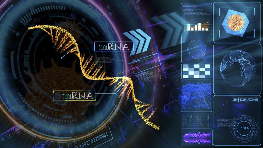
mRNA Technology: A Revolution in Medical Science
 By
By
mRNA technology revolutionises medicine by enabling rapid, cost-effective treatments, including vaccines, cancer therapies, and solutions for genetic and chronic diseases.
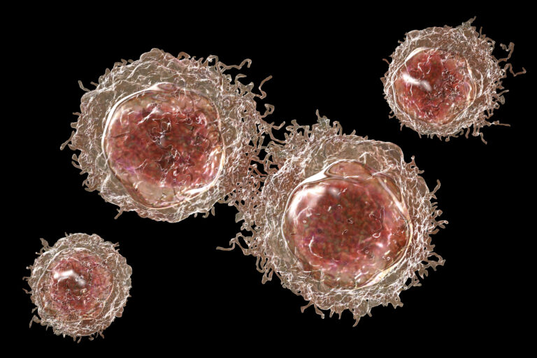
Cancer is a product of cellular changes that cause disease through the uncontrolled growth and division of cells. Over the past 150 years, many hypotheses have been proposed to explain the origin of cancer cells. These include the unregulated proliferation of cancer cells, metastasis and the histological classification of the cancer tissue (benign or malignant tumour).
In 1845, Rudolf Virchow found abnormal increases in white blood cells in some patients, and in 1847 identified the condition as a blood disease called leukämie (leukaemia). In 1857, Virchow was the first to identify chordoma, a tumour developed at the skull’s base. Also, in 1858, he presented that cancer cells are the body’s own cells.
Currently, the most accepted theory of cancer is based on the hysteron proteron of the somatic mutation theory (SMT). In this model, the first event (mutations) occurs after the cell has been transformed from a normal cell to a cancer cell via a process termed carcinogenesis. These mutations have increasingly been perceived as the causal event in the origin of the vast majority of cancers.
Currently, this model is challenged by a growing amount of experimental data and arguments that could either not be explained by the model or contradict this model.
One study of three subtypes of ependymoma (glioma) brain tumours found that one subtype carried an intrachromosomal translocation (a segment breaking off the chromosome and rejoining it at a different location), creating a new tumour-driving gene. The second subtype lacked tumour-driving mutations and had epigenetic modifications compared to the third subtype, which had neither gene mutations nor epigenetic aberrations.
Furthermore, the fact that normal tissues can display massive genetic changes, including changes in cancer-initiating and cancer-driving genes. Over the past few decades, several transfer experiments have demonstrated tumour-suppressing effects on normal cytoplasm. These include mitochondrial transfer, which has the ability to suppress tumour growth. This could be demonstrated – despite the presence of cancerous nuclear genomes – by showing the introduction of non-cancerous mitochondria into highly malignant breast cancer cells. This approach reversed the malignancy and down-regulated several oncogenic pathways, such as invasion and in vivo tumour growth.
Moreover, several non-genotoxic (non-mutagenic) carcinogens, including dichlorobenzene and chloroform, initiate cancer formation. Other oncological theories include oxidative phosphorylation, which results in cellular energy loss and highly impacts cancer formation.
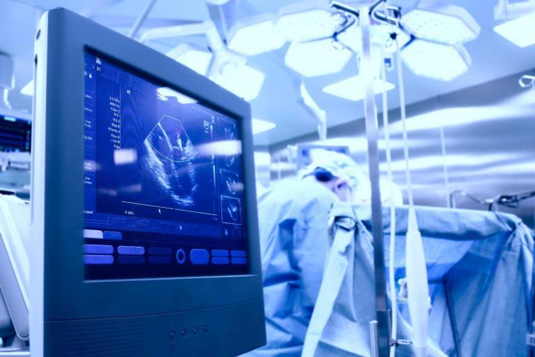
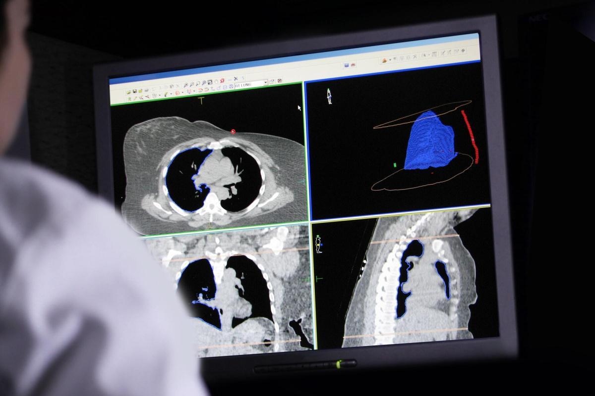
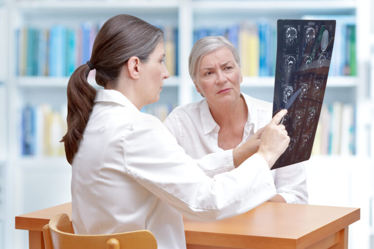
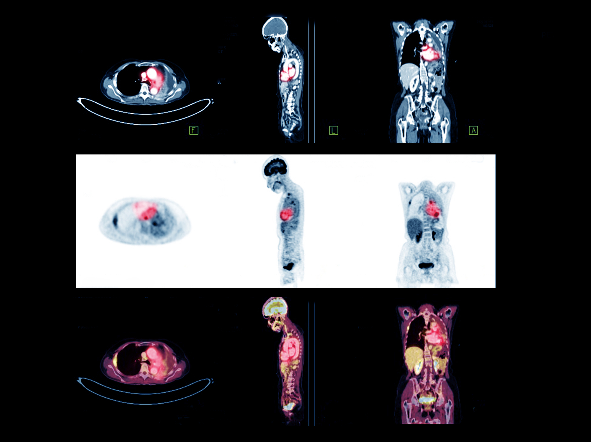
Currently, several imaging modalities are available to clinicians who diagnose, stage and treat human cancer: X-ray (plain film and computed tomography), ultrasound, magnetic resonance imaging, single-photon emission computed tomography, positron emission tomography and optical imaging. Of these, only four (MRI, CT, PET and SPECT) are capable of 3-D detection of cancer anywhere in the human body.
By
mRNA technology revolutionises medicine by enabling rapid, cost-effective treatments, including vaccines, cancer therapies, and solutions for genetic and chronic diseases.
 By
By
AI algorithms revolutionise tumour detection in medical imaging, enhancing precision, automating analysis, and supporting personalised cancer treatment through advanced PET/CT integration.
 By
By
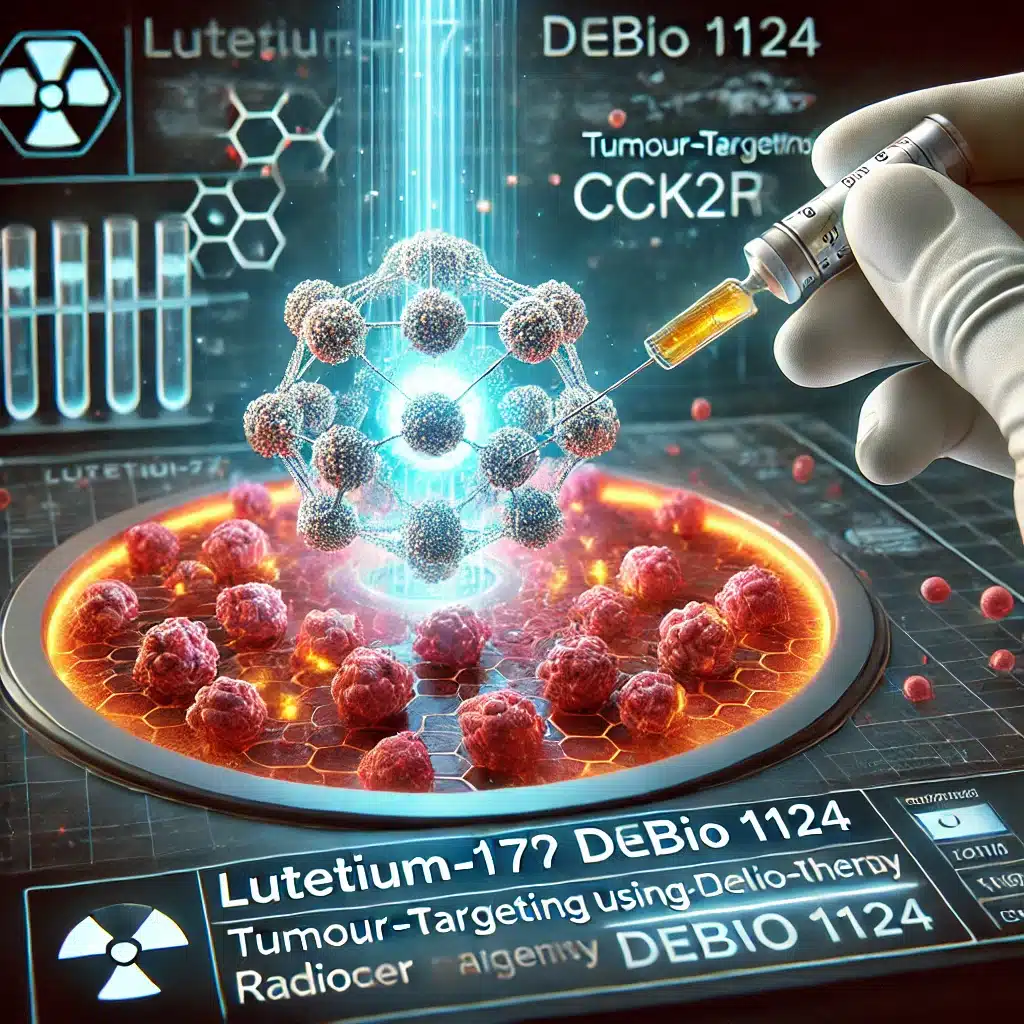
Lutetium-177 Debio 1124, a second-generation theranostic agent, selectively targets CCK2R-expressing tumours, offering precision radiotherapy and personalised oncology advancements.
 By
By
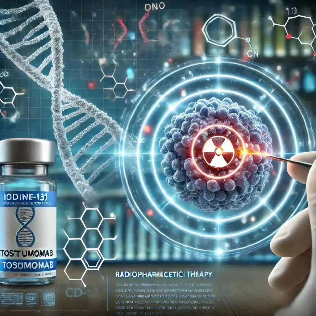
The rise and fall of Iodine-131 Tositumomab highlights challenges in balancing innovation, efficacy, infrastructure, and cost within radiopharmaceutical therapies.
 By
By
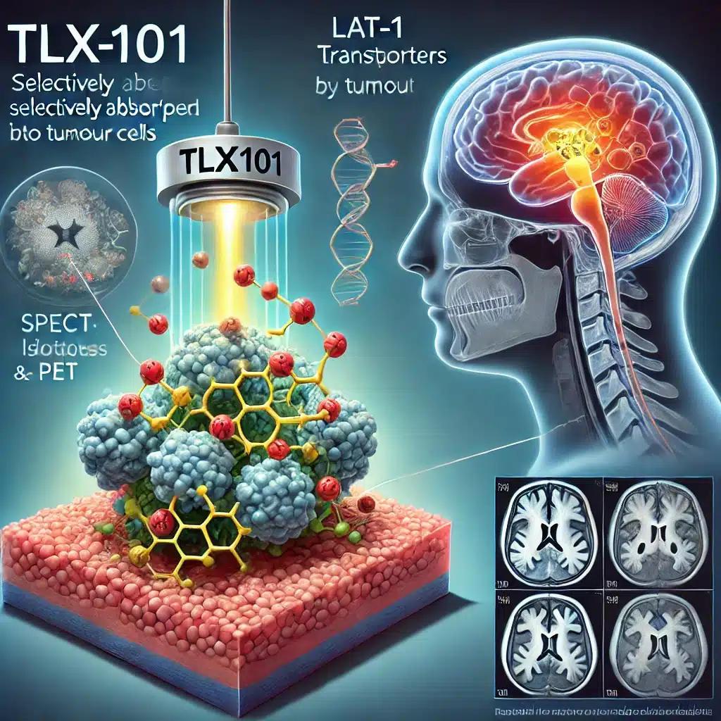
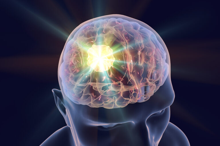
TLX101 is a revolutionary radiopharmaceutical targeting gliomas, offering dual imaging and therapy through tumour-selective uptake, enhancing treatment outcomes.
 By
By
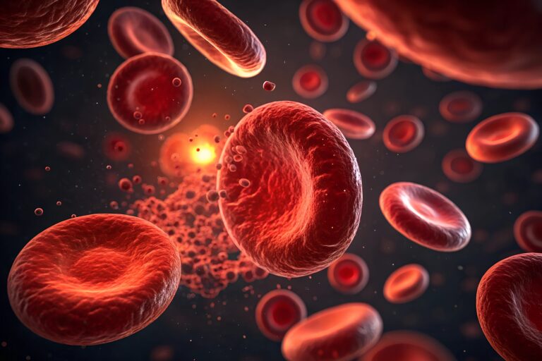
Imaging of blood types enhances medical diagnostics, ensuring accurate transfusions, organ compatibility, and advanced disease detection methods.
 By
By
Iodine-131 Omburtamab offers targeted radiation therapy, significantly improving survival in neuroblastoma patients with CNS and leptomeningeal metastasis.
 By
By
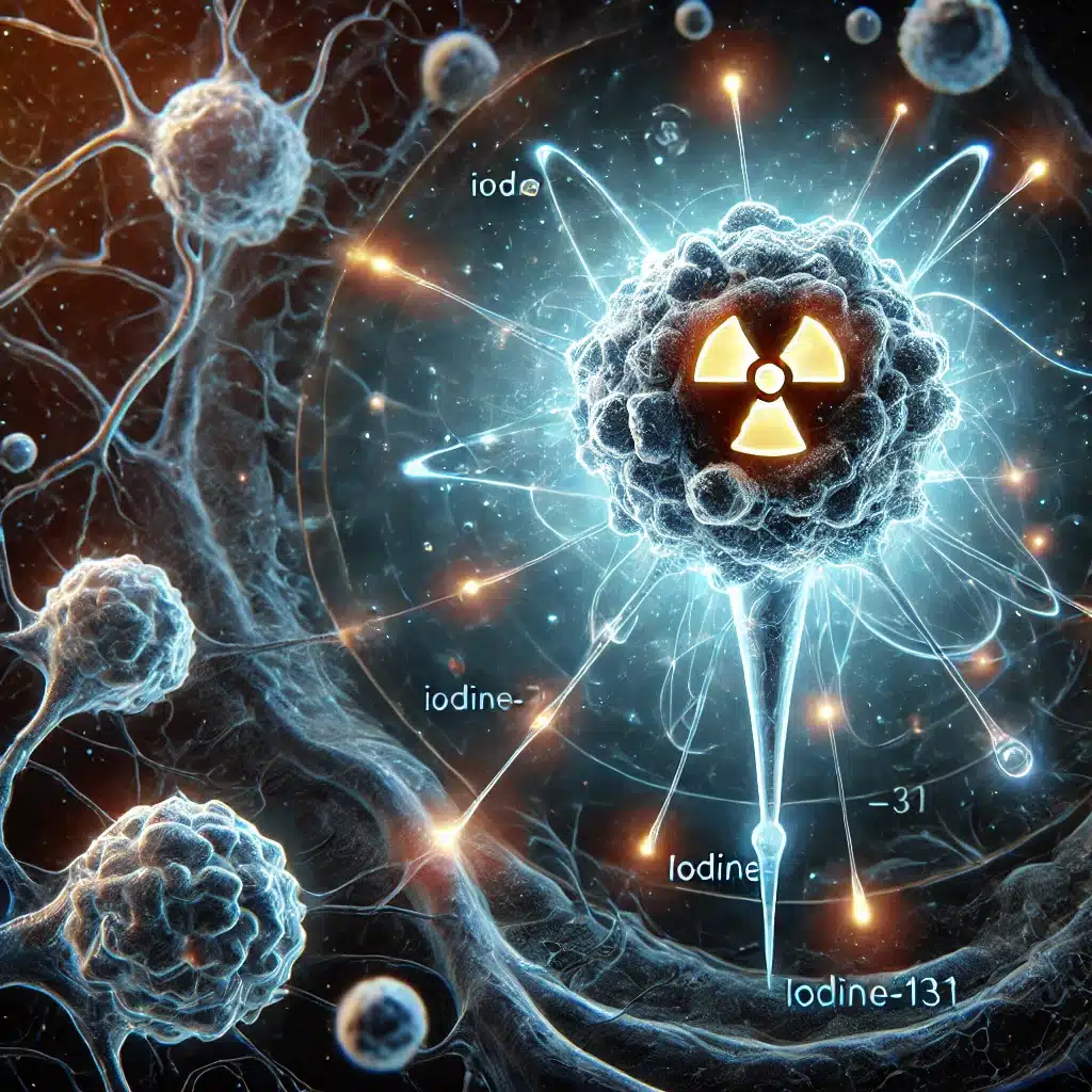
Iodine-131 naxitamab (¹³¹I-3F8) targets GD2-expressing cancers, offering precise radioimmunotherapy for neuroblastoma, melanoma, and small cell lung carcinoma.
 By
By
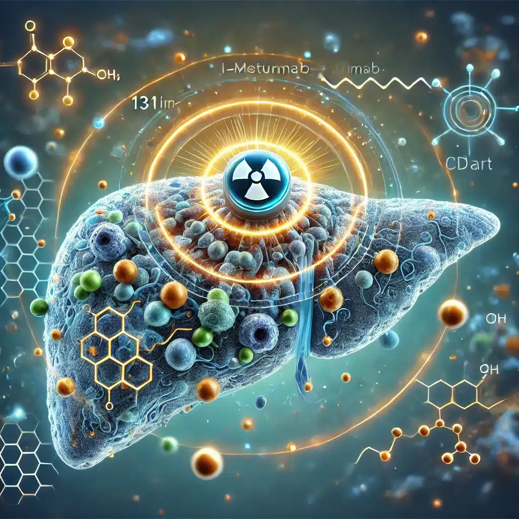
Iodine-131 Metuximab (Licartin) selectively targets CD147 receptors, delivering beta radiation to hepatocellular carcinoma cells, improving treatment precision significantly.
 By
By
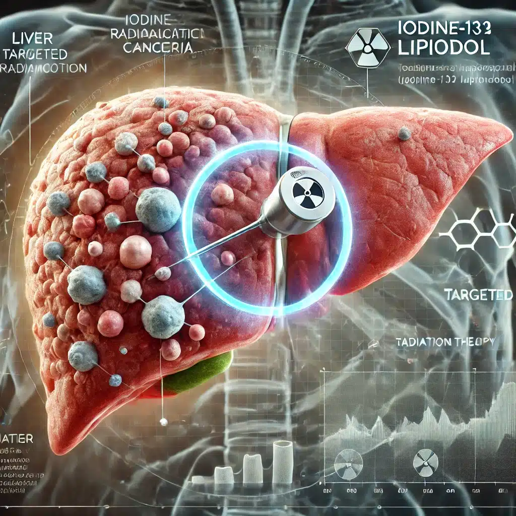
Iodine-131 Lipiodol has re-emerged as a promising therapy for hepatocellular carcinoma, particularly in non-resectable cases with portal vein thrombosis.
 By
By
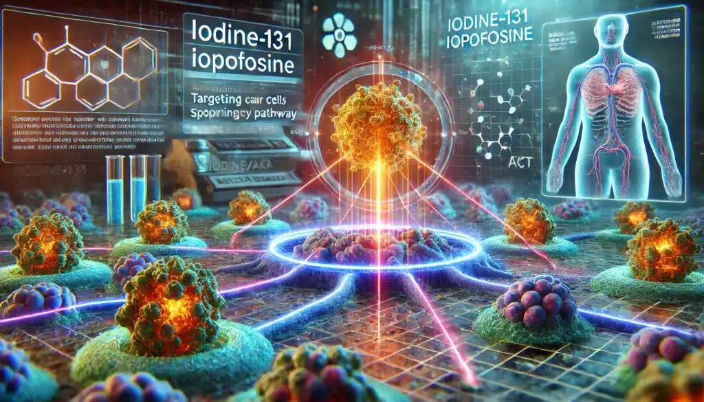
Iodine-131 Iopofosine selectively delivers cytotoxic radiation to malignant cells by targeting the PI3K/Akt pathway, offering promising cancer therapy advancements.
 By
By
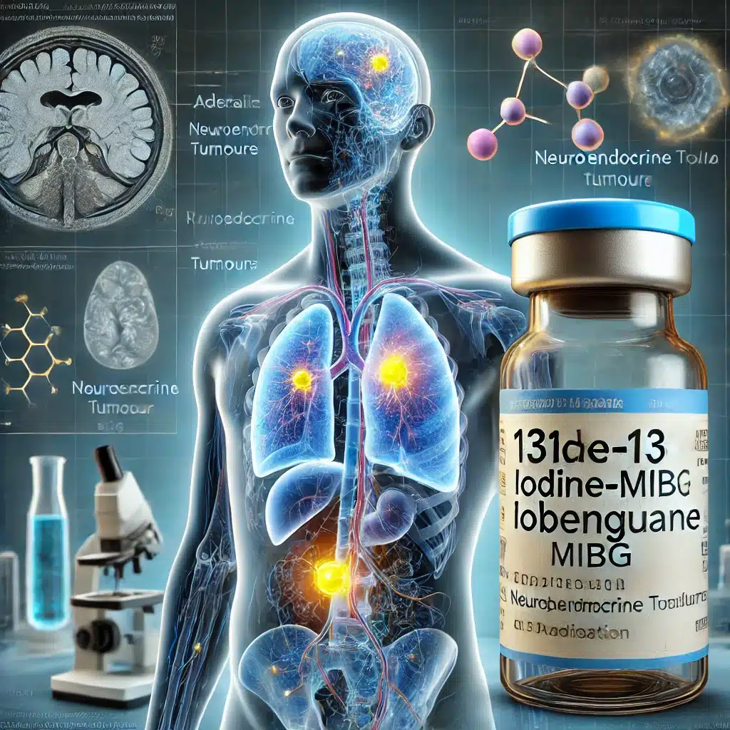
Iodine-131 Iobenguane revolutionises neuroendocrine tumour management by offering targeted imaging and therapy, significantly improving diagnosis, treatment, and patient outcomes.
 By
By
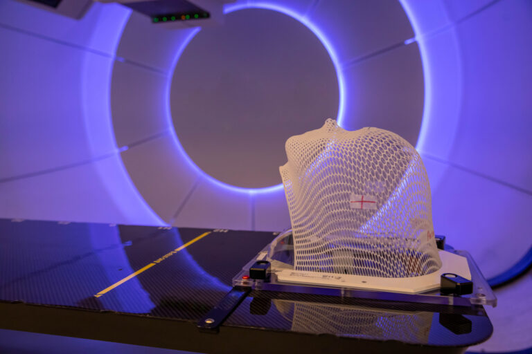
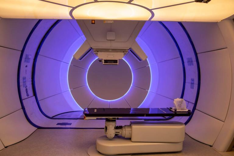
Advancements in Proton Beam Therapy have led to better cancer treatments with fewer side effects.
 By
By
Proton therapy in cancer treatment offers precise targeting, reducing damage to healthy tissues and improving outcomes.
 By
By
Blastoma tumours require advanced imaging techniques like MRI, CT, PET, and ultrasound for accurate diagnosis and staging.
 By
By
Radionuclide Therapy Effects include potential organ toxicity, requiring careful monitoring to manage patient outcomes effectively.
 By
By
Ultrasound plays a crucial role in cancer detection, offering a non-invasive, accessible, and effective tool for early diagnosis. Image for illustration only. People depicted are models.
 By
By
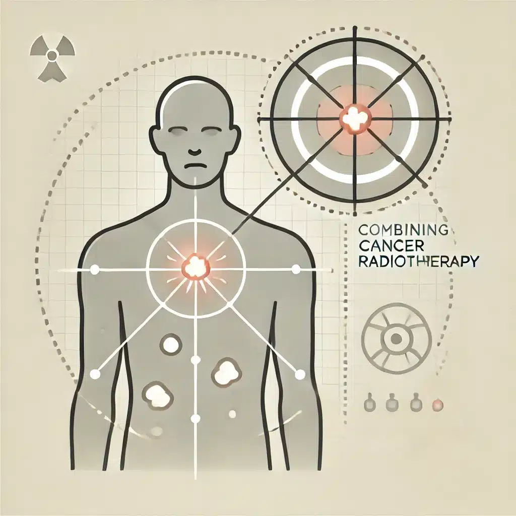
Cancer radiotheranostics combines targeted radiotherapy and diagnostic imaging to provide personalised, precise, and effective cancer treatment.
 By
By
This article explores cancer treatment advancements, tumour biology complexities, and the critical role of palliative care in oncology.
 By
By
Adverse effects of radionuclide therapy include fatigue, nausea, myelosuppression, renal toxicity, and secondary malignancies. Image for illustration only. Person depicted is a model.
 By
By
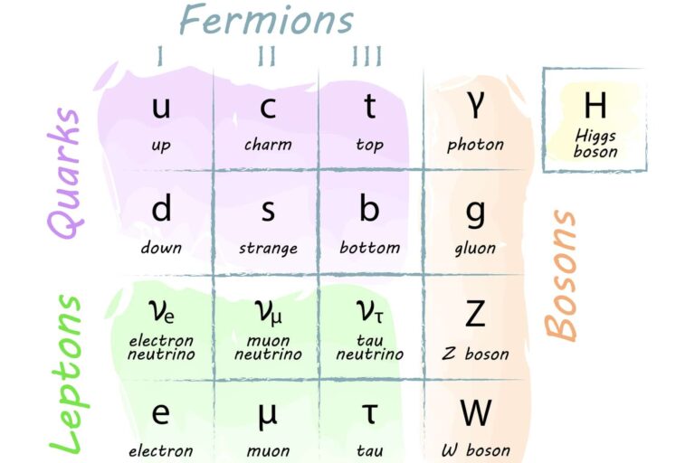
Fundamental particles form the essential building blocks of matter, underlying all physical phenomena and forces in the universe.
 By
By
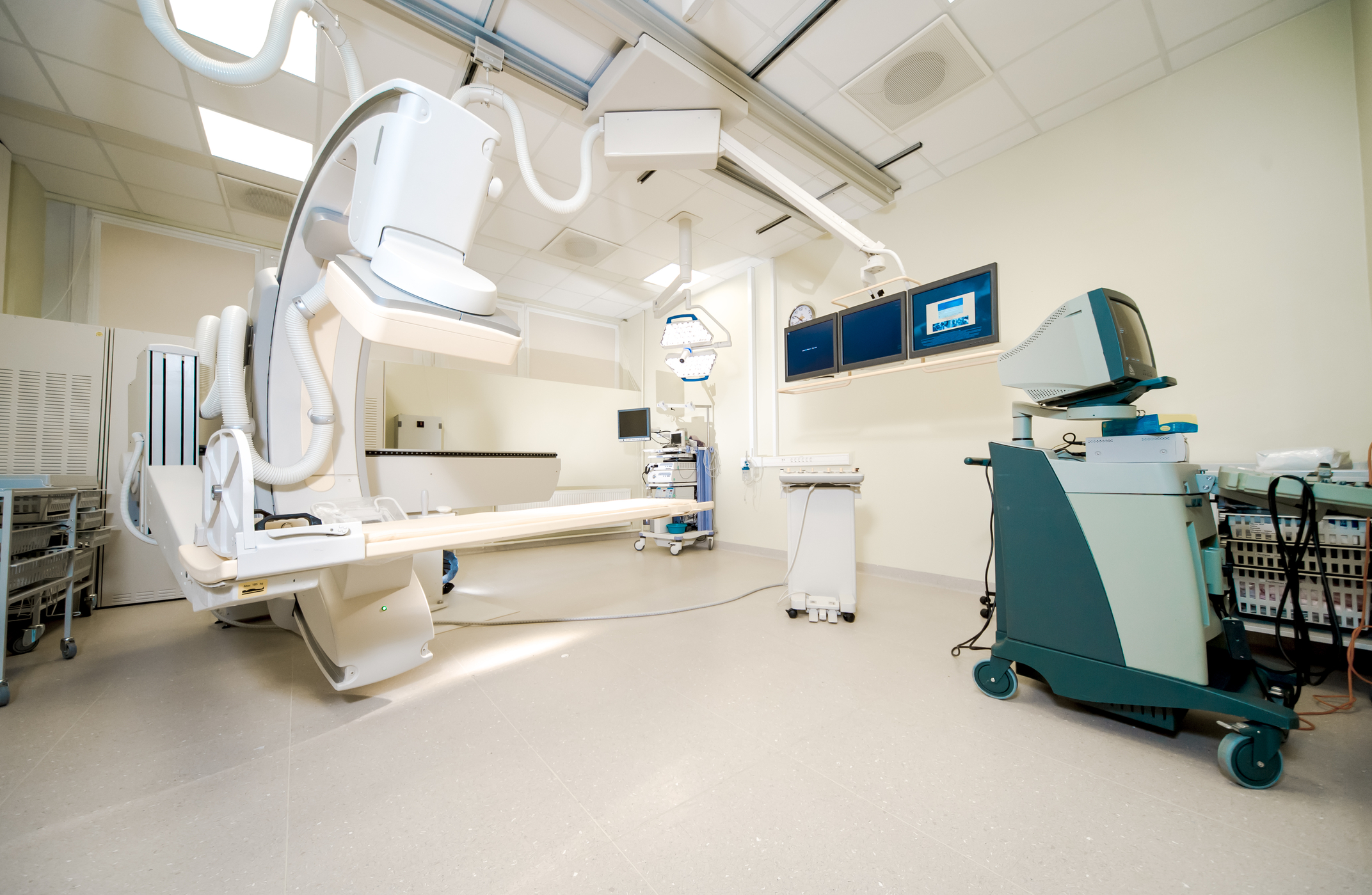
Advancements in medical imaging technology are transforming cancer diagnosis, enabling more precise treatment planning and better patient outcomes. Image for illustration only. People depicted are models.
 By
By
Auger electrons, crucial in surface science and medical physics, enable detailed material characterisation and targeted cancer therapies.
 By
By
This article examines the cancer risks associated with radionuclide administration in medical treatments and strategies for mitigation.
 By
By
Targeted Radionuclide Therapy for cancer delivers radioactive isotopes directly to tumour cells, maximising therapeutic effectiveness.
 By
By
Radiotheranostics combines diagnostic imaging and targeted radiotherapy, using radiopharmaceuticals for personalised, precise cancer treatment and improved outcomes.
 By
By
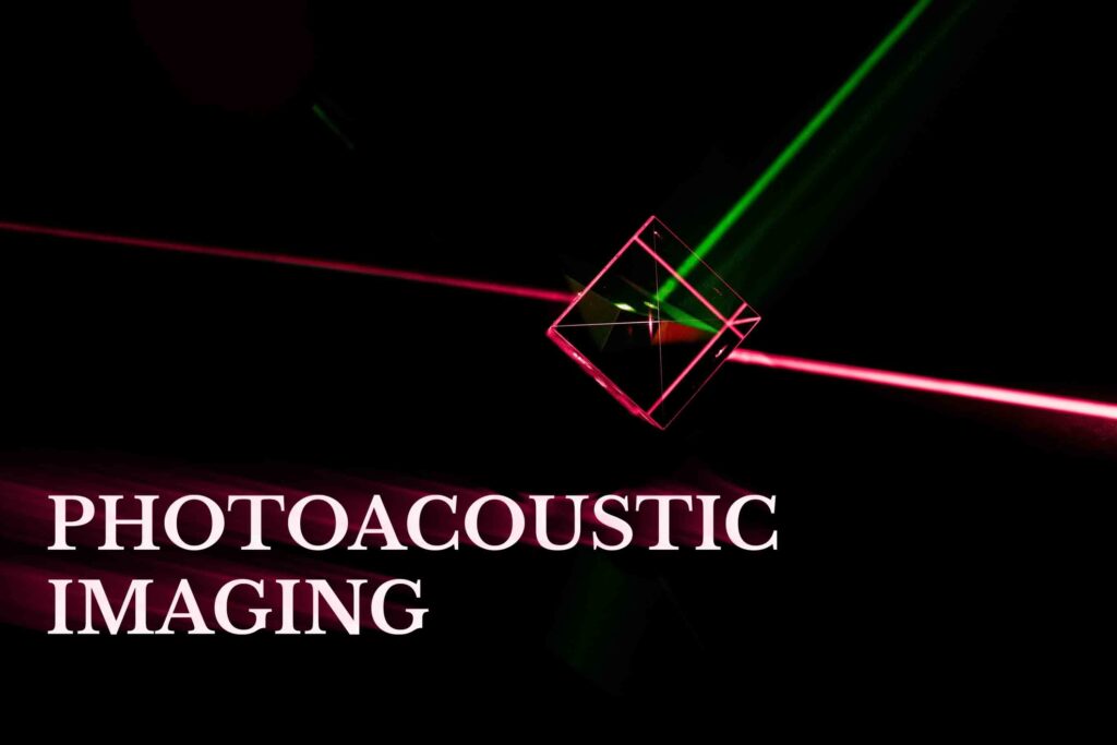
Photoacoustic imaging combines optical and ultrasound techniques, delivering high-resolution, non-invasive visualisations of biological tissues for enhanced diagnostics.
 By
By
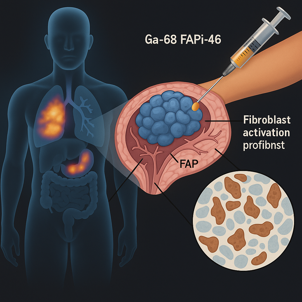
Advancing cancer diagnostics, the study evaluates Ga-68 FAPi-46 PET imaging’s potential to map FAP expression non-invasively in solid tumours.
 By
By
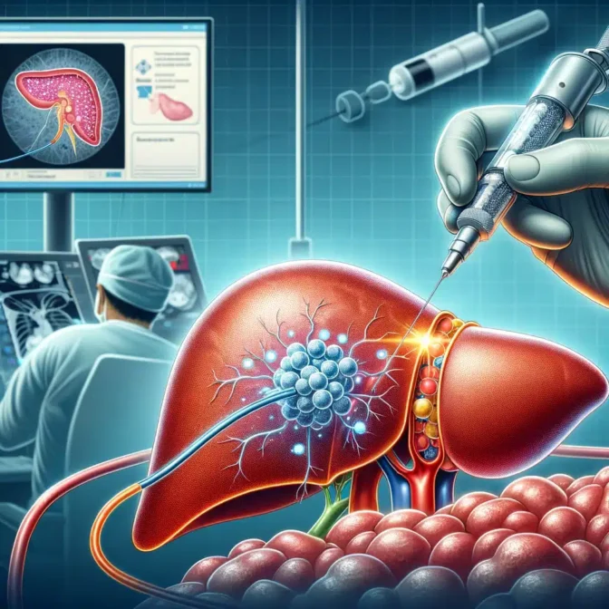
Selective Internal Radiotherapy (SIRT) delivers targeted radiation to liver tumors, providing effective treatment with minimal damage to surrounding healthy tissue.
 By
By
Targeted Alpha Radionuclide Therapy precisely delivers potent alpha particles to tumors, maximising efficacy while sparing healthy tissues.