Revolution in the Spinal Cord: How New Imaging Tech is Transforming Diagnosis and Care
 By
By
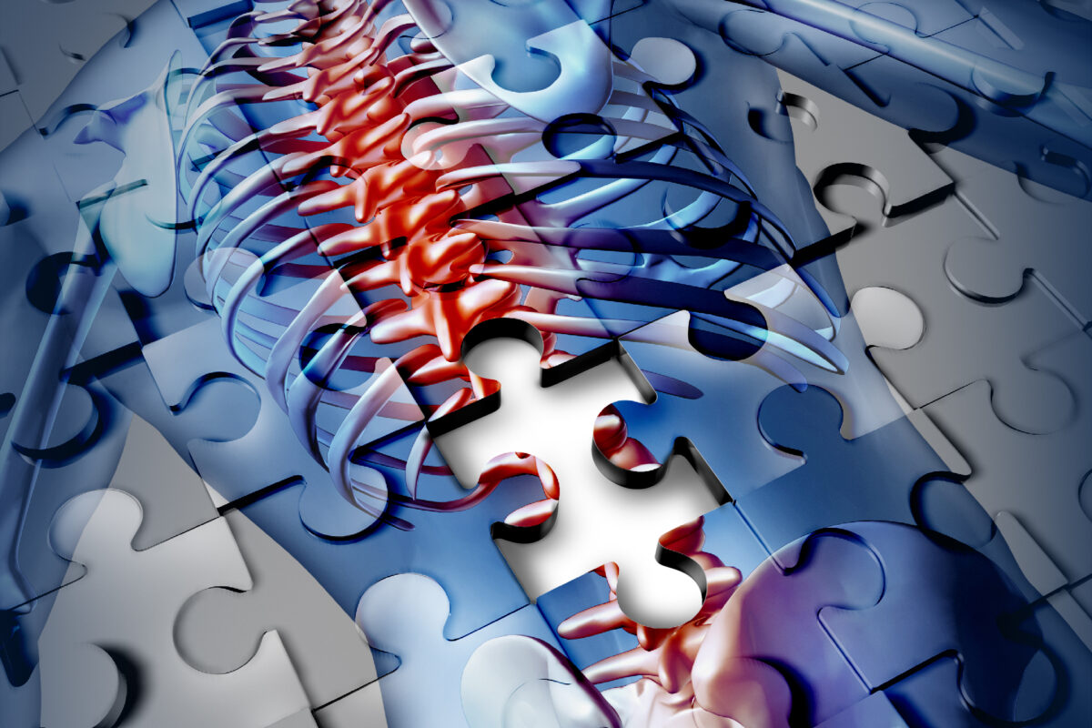
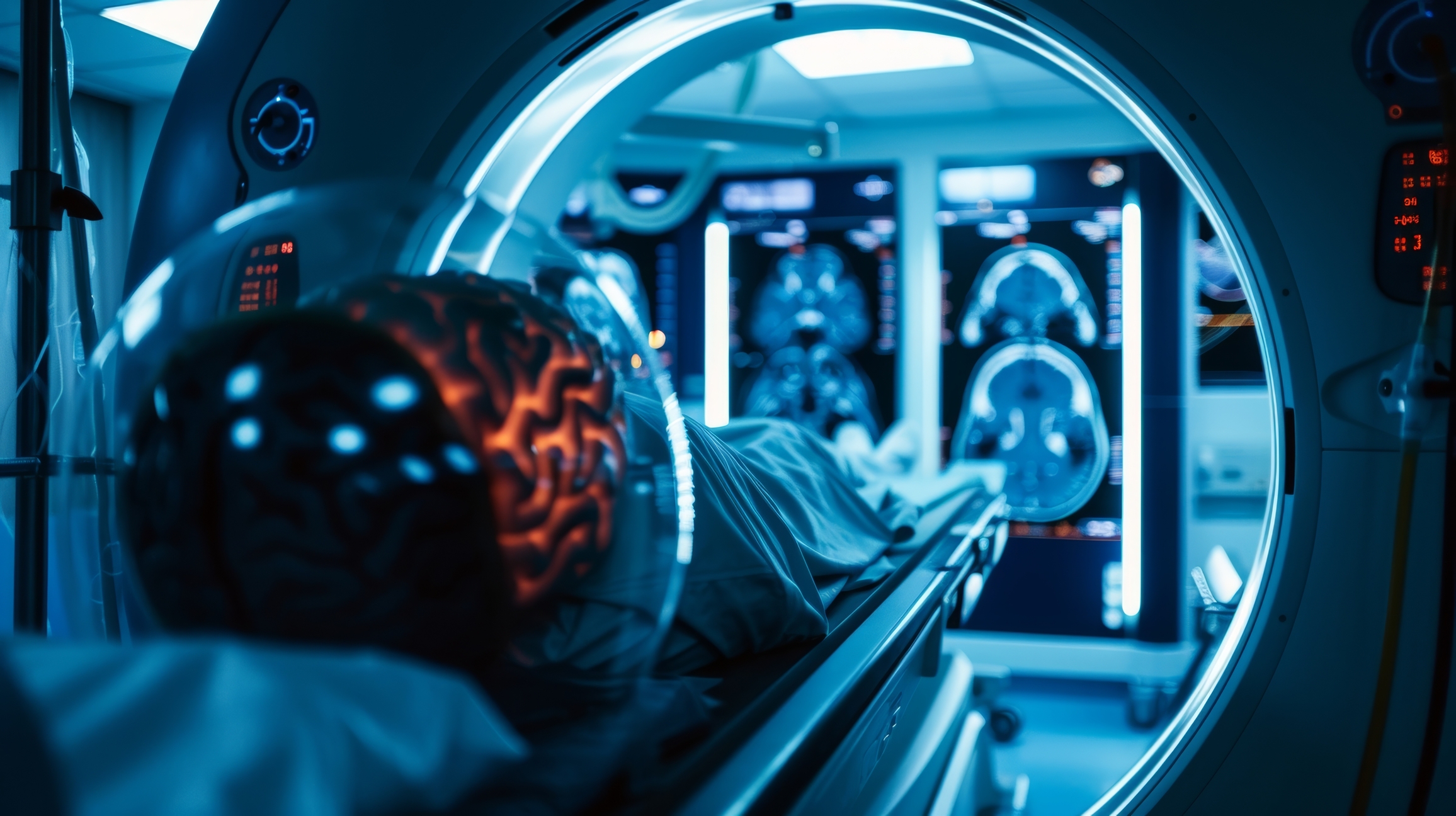
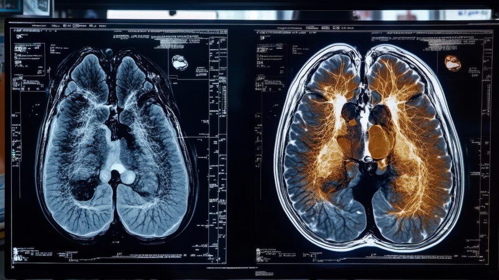
Discover the latest developments in spinal cord imaging, including ultra-high-field MRI and artificial intelligence enhancements.
By
Discover the latest developments in spinal cord imaging, including ultra-high-field MRI and artificial intelligence enhancements.
 By
By
Find out what causes the loud noise from MRI scanners. MRI scanner noise explained for better patient understanding and comfort.
 By
By
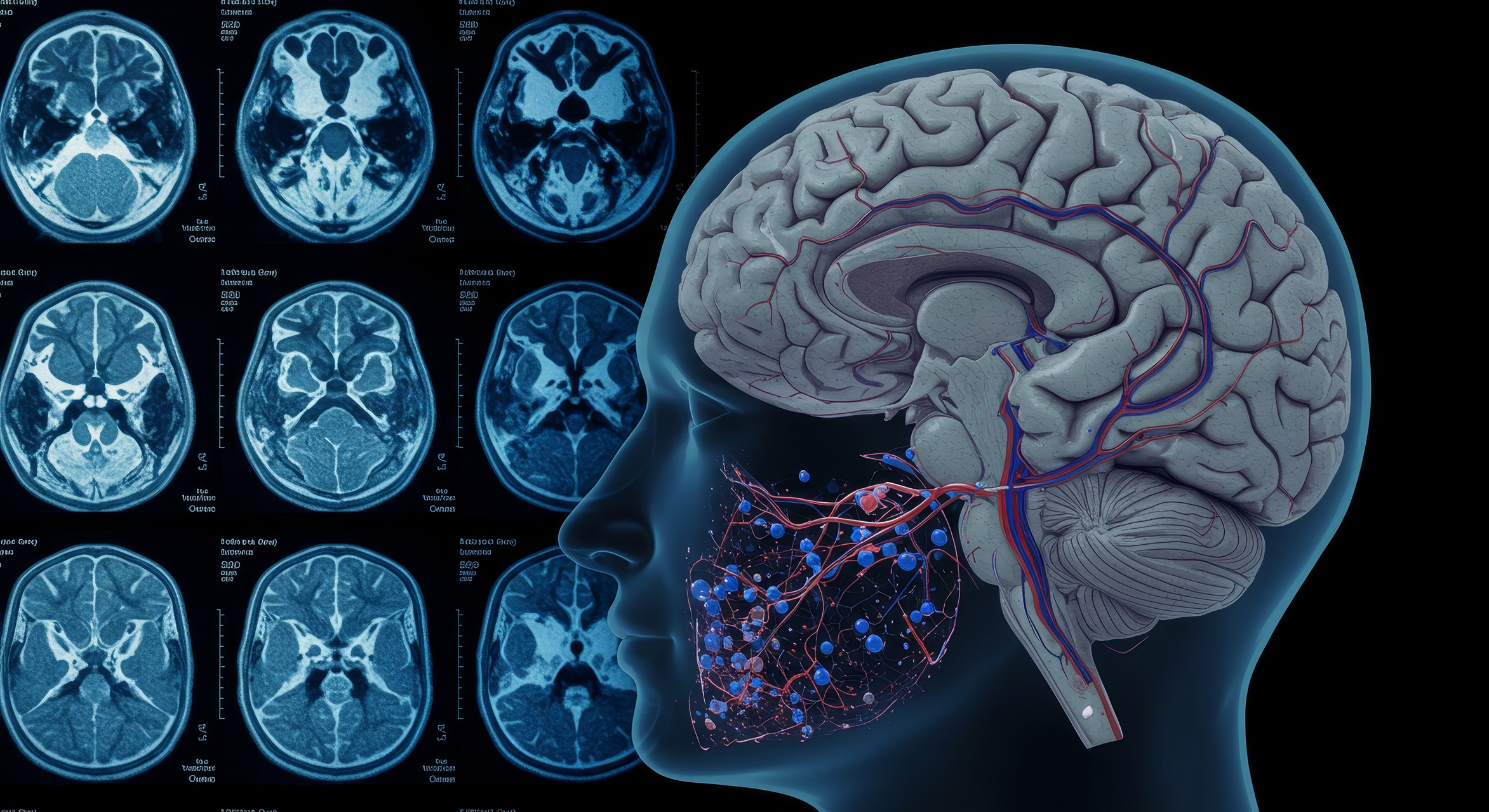
Learn how functional MRI technology is used to assess brain injury. Understand the complexities of mTBI symptoms and recovery.
 By
By
Explore the link between student mental health and brain imaging. Discover how stress impacts students’ academic success.
 By
By
Uncover the facts about MRI safety and metal objects. Find out how these items can become dangerous projectiles in MRI environments.
 By
By
Discover the principles behind magnetic resonance theory and how it has advanced MRI as a vital tool in modern diagnostic imaging.
 By
By
Find out how AI in MRI scanning improves workflow efficiency by enhancing image quality and minimising contrast agent use.
 By
By
CEST MRI reveals molecular tissue details, enhancing early disease diagnosis without external contrast agents.
 By
By
Compressed Sensing in MRI transforms patient diagnostics by drastically reducing scan times without compromising image quality.
 By
By
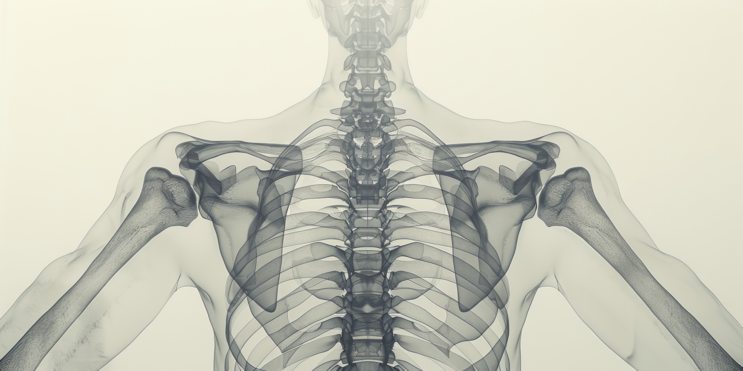
Medical imaging of the human skeleton enables accurate diagnosis, treatment, and monitoring of diverse bone and joint conditions.
 By
By
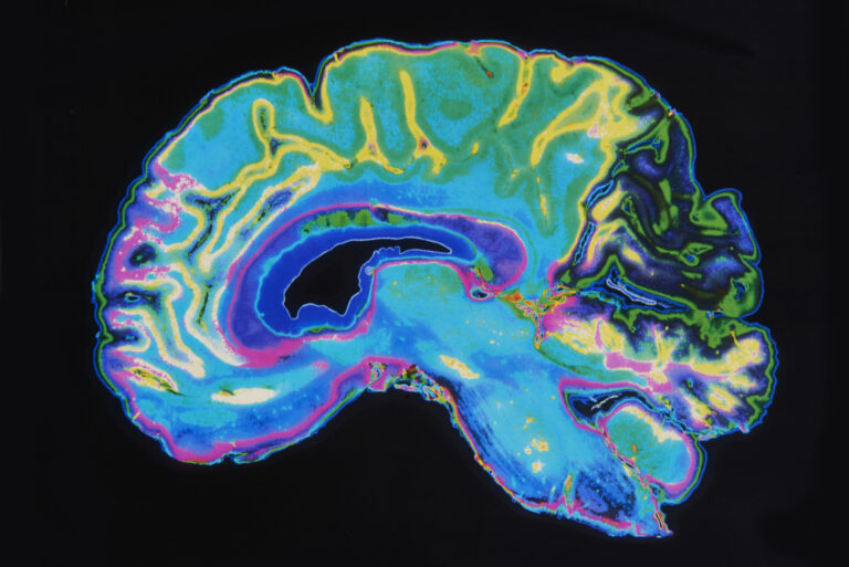
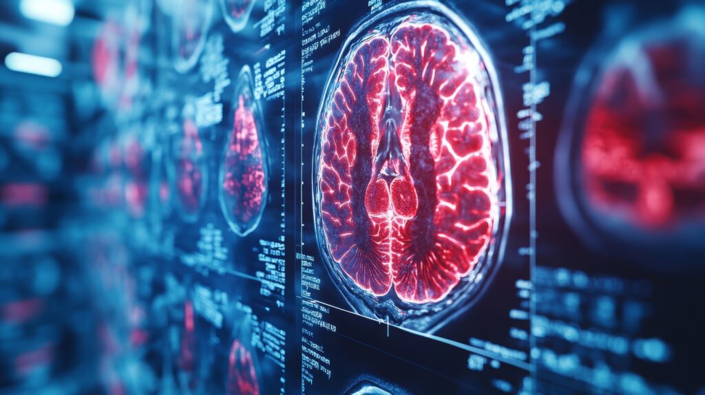
A useful brain imaging technique uses functional magnetic resonance imaging to analyse metabolic changes such as blood oxygenation.
 By
By
Medical imaging modalities, including MRI, CT, and ultrasound, facilitate accurate diagnoses and treatments.