Carbon-14 Radiolabelling and the Role of Beta Particles in Molecular Stability
 By
By
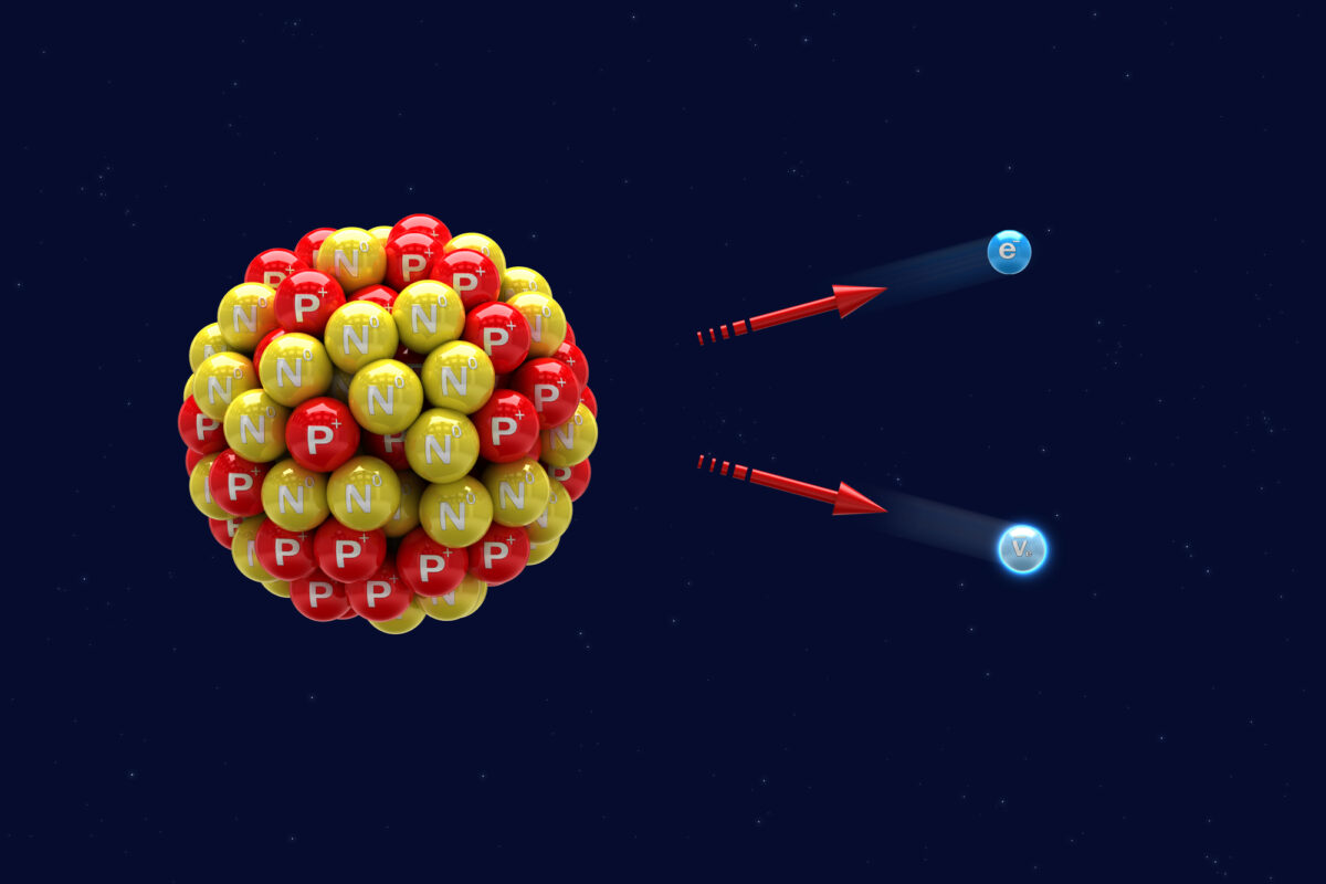
Learn how Carbon-14 beta particles enhance biochemical research, allowing scientists to track drugs through metabolic pathways.
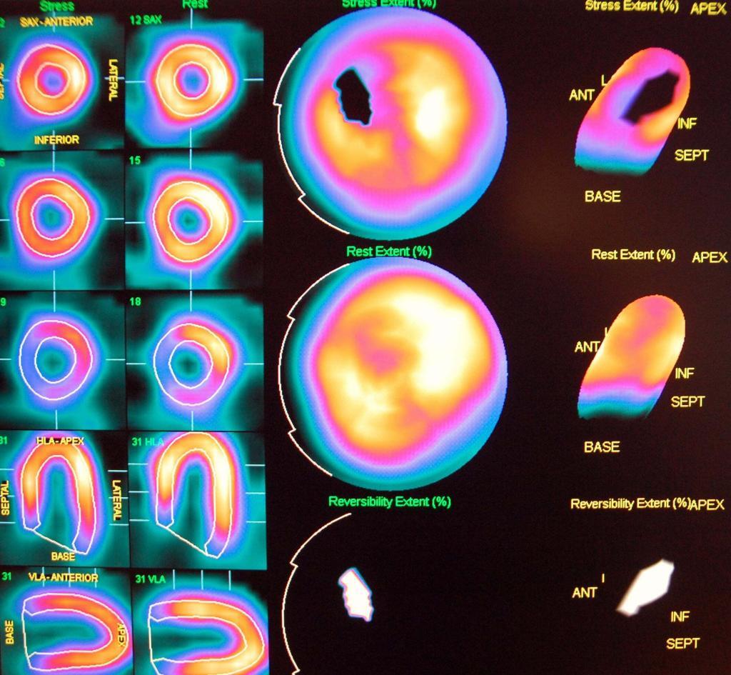
Radionuclide Imaging is fundamental to nuclear medicine, and the first radionuclide studies used [131I]-sodium iodide for diagnosing and treating thyroid cancer. Therefore, nuclear medicine consists of radionuclide imaging and radionuclide therapy and is mostly done on patients requiring the former. However, both radionuclide imaging and therapeutic procedures are increasing because of personalised medicine used in the design of patient treatment plans for the management of cancer.
To perform imaging, the radiopharmaceutical is administered to the patient and the radiation emitted by the radiotracer is detected by a series of gamma cameras around the patient. Radionuclide imaging in the clinical setting can be divided into a positron and single-photon imaging.
The radioisotopes used for single-photon imaging emit gamma rays in the energy range of approximately 75-360 keV. These radionuclides include technetium-99m (the metastable nuclear isomer of technetium-99), iodine-131, thallium-201 (myocardial perfusion imaging), indium-111 (radiolabelled red cells, platelets and leukocytes), gallium-67 (detect the presence of acute inflammatory lesions and various malignancies including Hodgkin’s disease, lymphomas and bronchogenic carcinomas).
Several gamma cameras are used to detect these radionuclides and can perform regional, single-photon emission computed tomography (SPECT), and whole-body imaging. The other common positron-emitting radiotracers include fluorine-18, carbon-11, nitrogen-13 and oxygen-15. These can include radionuclide imaging because the positron travels a short distance through the tissue and interacts with a free electron.
At this point, the positron and electron annihilate each other, generating two photons (two gamma rays) with energies of 511 keV in opposite directions. The gamma cameras around the body detect the coincidence radiation. However, positron emission tomography is more accurate for scattering the radiation – by absorption in the body – than SPECT imaging and also provides higher-resolution lesion detection.
The advancements in radionuclide therapy and imaging involve the combination of PET and computed tomography (CT) scanners. Therefore, the hybrid PET/CT and SPECT/CT scanners can obtain high-resolution anatomical information from CT imaging and biological information from PET and SPECT imaging.
home »
By
Learn how Carbon-14 beta particles enhance biochemical research, allowing scientists to track drugs through metabolic pathways.
 By
By
Zirconium-89 molecular imaging revolutionises diagnostics by enabling precise tumour localisation and tracking antibody-based therapies effectively.
 By
By
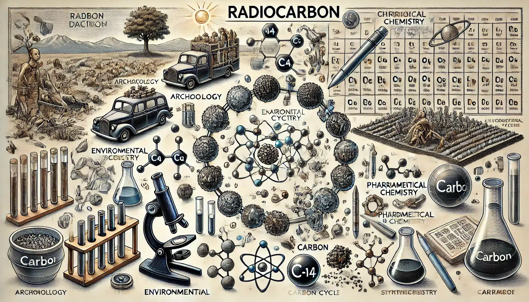
Radiocarbon chemistry enables precise insights into carbon cycles, archaeological dating, drug development, and environmental impact analysis worldwide.
 By
By
Carbon-14 offers distinct radiolabelling benefits, including a long half-life, minimal molecular alteration, and extensive applications in research.
 By
By
The fine-structure constant, denoted by α, is fundamental in understanding electromagnetic interactions between charged particles in physics.
 By
By
The atomic mass unit (amu) allows scientists to measure atomic and molecular masses with remarkable precision.
 By
By
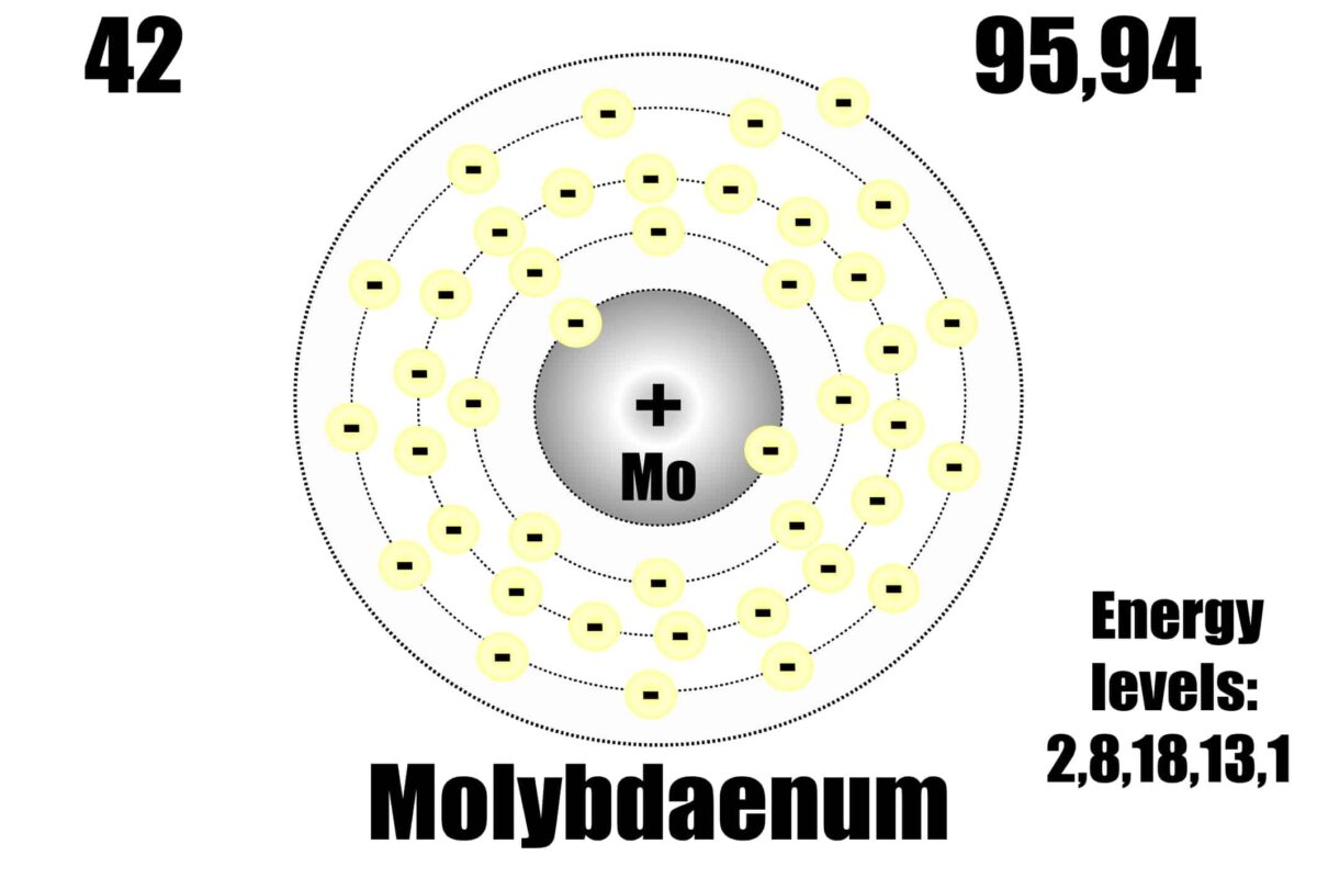
The nuclear shell model explains nucleon arrangement in discrete energy levels, leading to nuclear stability and magic numbers.
 By
By
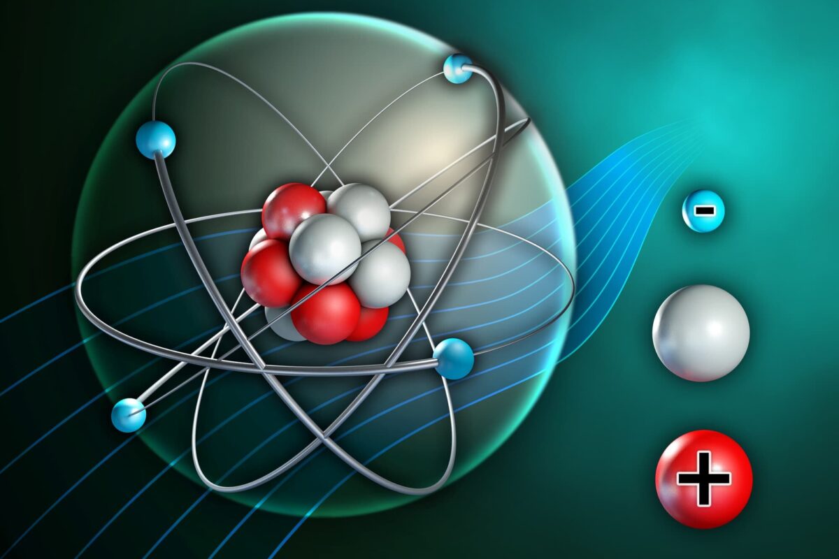
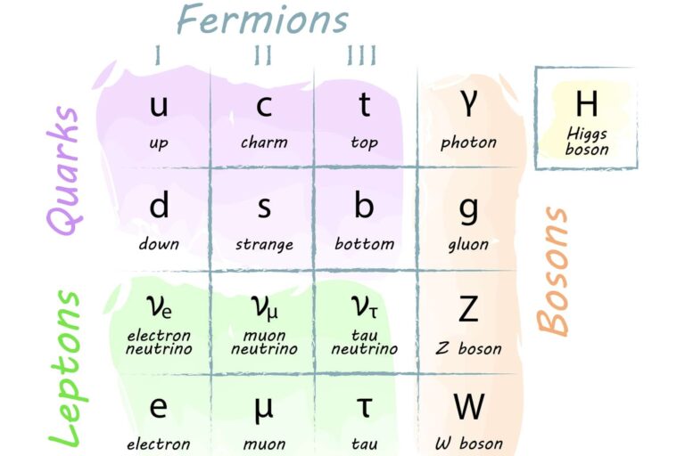
Fundamental particles form the essential building blocks of matter, underlying all physical phenomena and forces in the universe.
 By
By
Electron capture transforms a proton into a neutron by absorbing an inner electron, significantly altering the atomic nucleus.
 By
By
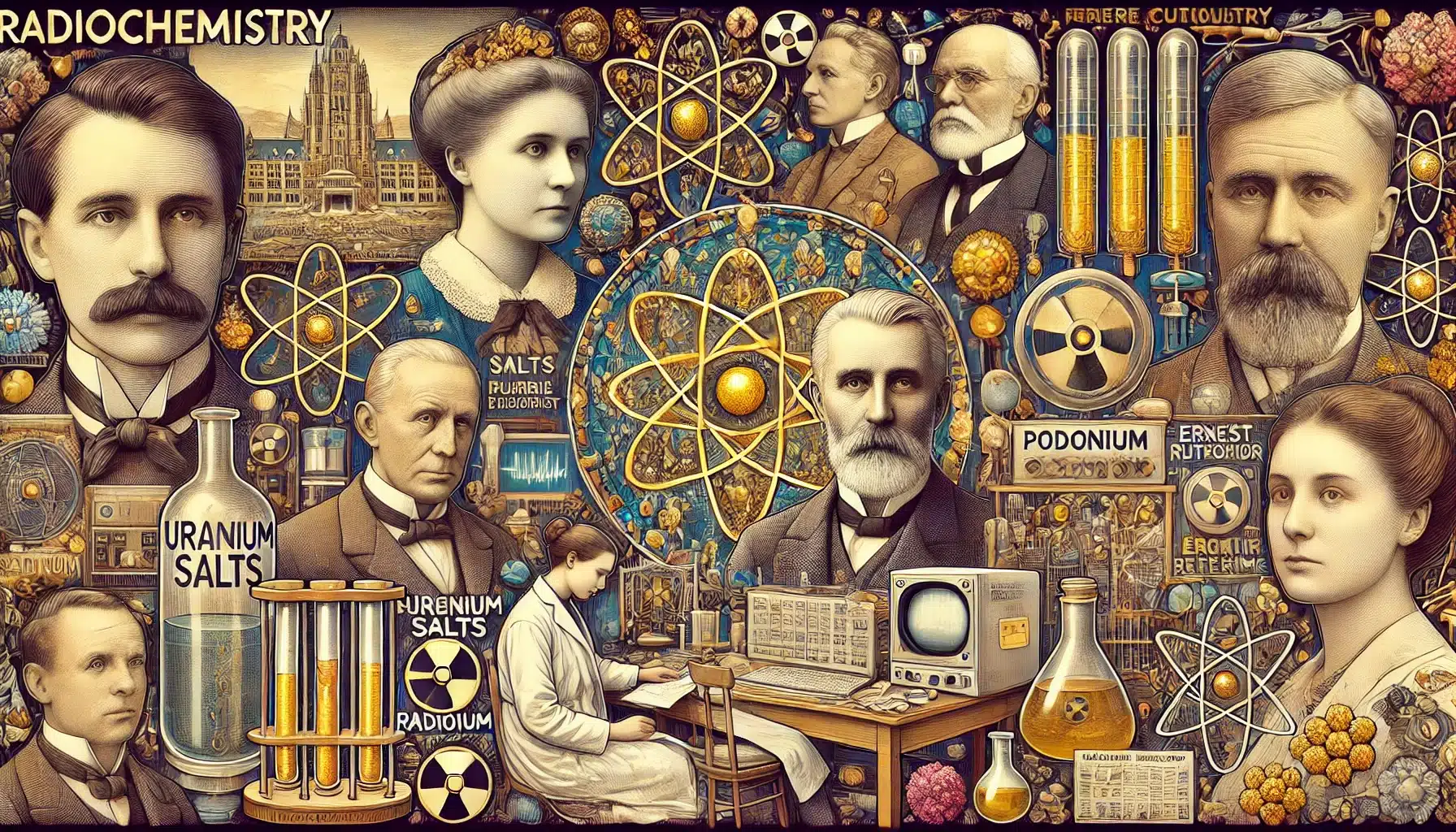
History of Radiochemistry chronicles its evolution from the discovery of radioactivity to modern applications in science and medicine.
 By
By
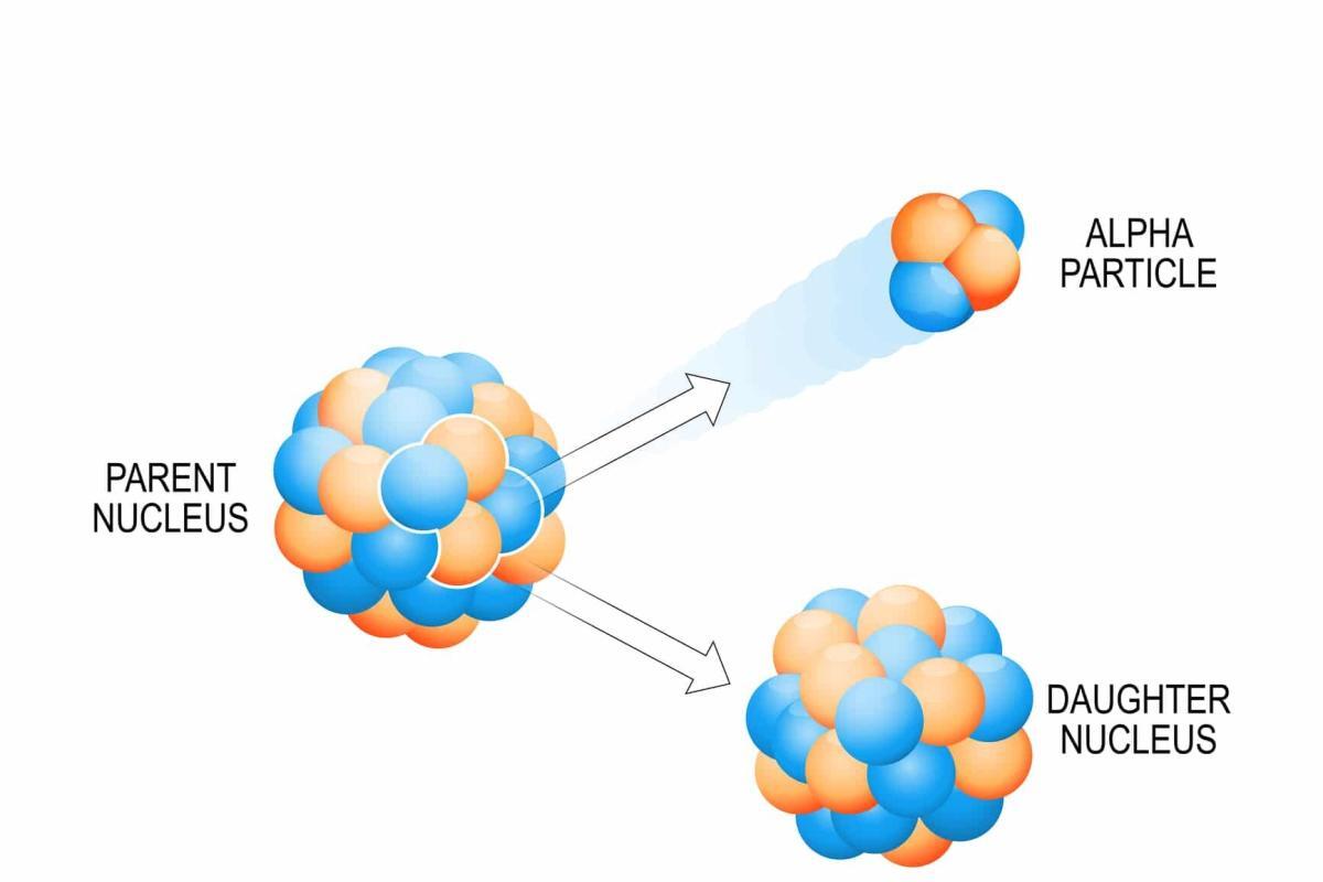
Alpha particles, with high ionising power and low penetration, significantly impact biological tissues when internalised in the body.
 By
By
SPECT imaging combines nuclear medicine with computed tomography to deliver functional insights, revolutionising diagnosis and treatment across cardiology, neurology, and oncology.
 By
By
Folate Receptor Targeting Agents revolutionise cancer diagnosis and treatment with Fluorine-18 and Rhenium-188 labelling.
 By
By
Indium-111 chloride effectively aids infection and inflammation imaging by labelling WBCs, enabling precise issue localisation and severity assessment.
 By
By
Imaging agents can be used to evaluate organ function, detect cancer, measure blood flow and follow metabolic processes.