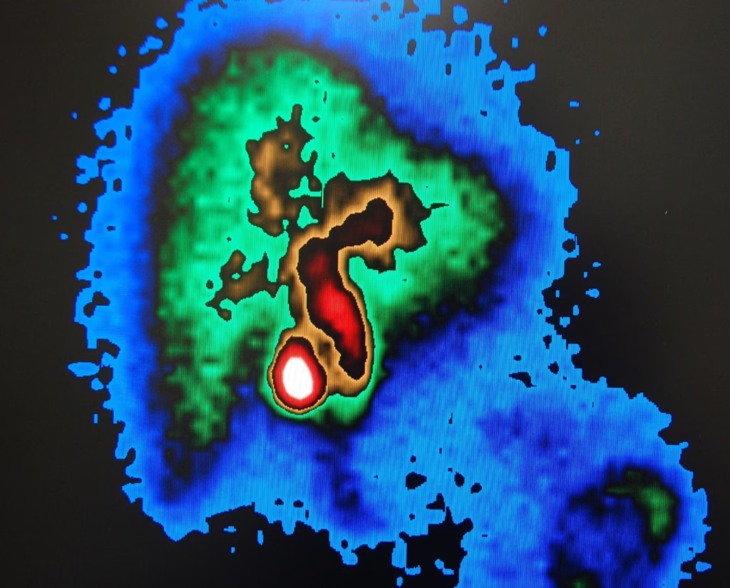
Summary: In today’s dermatological landscape, the integration of cutting-edge imaging technologies has transformed how clinicians diagnose, monitor, and treat various skin conditions. Through non-invasive scans and artificial intelligence-driven tools, specialists can now visualise the skin’s structure in unprecedented detail. This article looks into the world of advanced dermatological imaging, uncovering the methods that allow practitioners to refine diagnoses, improve patient outcomes, and detect dangerous skin diseases at their earliest stages.
Keywords: skin imaging techniques; non-invasive diagnostic tools; dermatology imaging advancements; high-resolution skin scans; AI-based dermatological analysis; early melanoma detection methods
Focus Keyword: advanced dermatological imaging
The Need for Advanced Imaging in Dermatology
Skin health is undergoing a radical transformation as cutting-edge medical imaging technologies redefine the way dermatologists and researchers assess and manage conditions. By using advanced dermatological imaging systems, experts can capture detailed insights into the human epidermis and dermis, unlocking previously hidden layers of information. The significance is huge: from identifying suspicious lesions to evaluating chronic skin conditions, these imaging modalities are providing a new lens for diagnostic accuracy and treatment planning.
Historically, dermatologists relied primarily on visual inspections with the naked eye, dermoscopy, and histopathological analysis of biopsied samples. Although these methods have long provided valuable insights, they have inherent limitations. For instance, standard visual examinations can fail to detect subtle skin abnormalities or resolve the intricate structure beneath the surface. Biopsies, while informative, require tissue sampling, which can be invasive, uncomfortable, and potentially scarring.
The advent of advanced dermatological imaging changes this equation. By using skin imaging techniques that present a clear, non-invasive view, it is now possible to identify subtle variations in skin morphology and pigment distribution that were not visible before. This shift reduces the need for unnecessary biopsies and streamlines the diagnostic process, leading to faster, more accurate decision-making.
Integrating Non-Invasive Diagnostic Tools
Incorporating non-invasive diagnostic tools into everyday clinical practice allows dermatologists to examine the skin’s deeper layers without physically penetrating the tissue. Techniques like confocal microscopy, optical coherence tomography (OCT), and multispectral imaging enable clinicians to visualise cellular-level details and differentiate between benign and malignant lesions with greater confidence.
These dermatology imaging advancements offer more than just patient comfort. They also enhance diagnostic accuracy by improving lesion characterisation and helping clinicians distinguish between conditions that may appear similar on the surface. This is where high-resolution skin scans excel: by magnifying and clarifying microscopic details, dermatologists can formulate targeted treatment plans that lead to improved patient outcomes.
Key Imaging Modalities and Techniques
Dermoscopy: For years, dermoscopy has served as a gateway to enhanced visual inspection. By magnifying the skin’s surface, it helps dermatologists identify suspicious pigmented lesions. However, newer imaging methods push the boundaries even further.
Reflectance Confocal Microscopy (RCM): RCM provides cellular-level resolution, allowing clinicians to observe skin layers with almost histological detail. It stands out among skin imaging techniques for its capacity to differentiate between benign and malignant lesions without the need for immediate biopsy. Using RCM, practitioners can visualise individual melanocytes, track their shape and distribution, and thereby improve the specificity of diagnosis.
Optical Coherence Tomography (OCT): OCT works by reflecting infrared light off tissue structures to produce cross-sectional images of the skin. It has proven particularly beneficial for diagnosing non-melanoma skin cancers and inflammatory conditions. OCT’s ability to generate high-resolution images in real-time makes it a cornerstone of non-invasive diagnostic tools.
Multispectral and High-Resolution Imaging
Multispectral Imaging: By using multiple wavelengths of light, multispectral imaging analyses how the skin absorbs and reflects certain frequencies. The result is a clearer understanding of underlying structures, pigmented lesions, and vascularisation. This approach not only enhances the detection of atypical lesions but also guides treatment planning.
High-Resolution Skin Scans: Another pillar of dermatology imaging advancements is the deployment of high-resolution skin scans. These scans, obtained through cutting-edge cameras and scanning systems, offer exceptionally detailed images of the skin’s texture, pores, and vascular networks. In combination with other modalities, they provide a comprehensive data set that elevates the diagnostic process.
Artificial Intelligence and Machine Learning in Dermatology
Human interpretation of imaging data can be subjective and influenced by experience or time constraints. This is where AI-based dermatological analysis comes into play, significantly enhancing the accuracy and speed of image interpretation. Machine learning algorithms, trained on vast datasets, can rapidly distinguish between benign and malignant lesions, classify skin conditions, and even predict a lesion’s risk level.
Advanced dermatological imaging combined with AI capabilities reduces the burden on clinicians, offering preliminary assessments that can guide further examination. By integrating these tools into their workflow, dermatologists gain a second pair of “eyes” – a digital assistant that never tires, always remains objective, and constantly learns from new data.
Augmenting Human Expertise
It is crucial to recognise that AI does not replace the dermatologist. Instead, it complements clinical expertise by filtering through images rapidly and identifying patterns that may be imperceptible to the human eye. This synergy enables dermatologists to focus on complex cases, invest more time in patient communication, and continuously refine diagnostic accuracy.
Incorporating AI-based dermatological analysis into routine practice provides a more comprehensive approach to patient care. By harnessing technology’s strengths and combining it with human clinical acumen, the result is a significant boost to the standard of dermatological healthcare.
Identifying Early Signs with Advanced Dermatological Imaging
Skin cancer, and melanoma in particular, poses a serious risk to global populations. Fortunately, early melanoma detection methods are improving as advanced dermatological imaging makes subtle changes in skin lesions easier to spot. By capturing minute differences in size, shape, and colour, these imaging technologies alert clinicians to potential melanomas before they become dangerous.
Comprehensive skin imaging techniques, from RCM to OCT, allow for close examination of atypical moles. This ensures that changes are caught at their earliest stages. Combined with non-invasive diagnostic tools, these imaging methods empower dermatologists to intervene sooner, increasing the likelihood of successful treatment.
Empowering Patients Through Awareness
Part of effective early detection involves patient awareness. Patients can monitor their own skin changes with the guidance of home-based imaging devices, smartphone apps, and tele-dermatology services. While these tools are not a substitute for professional assessment, they encourage individuals to report suspicious lesions earlier.
This proactive approach, guided by physicians, leverages early melanoma detection methods that rely on detailed images and robust datasets. Ultimately, patients become active participants in their own care, fostering better communication and trust between them and their dermatologists.
Dermatology Imaging Advancements Shaping the Future
The future of dermatology imaging advancements lies in integrative approaches. By combining advanced dermatological imaging data with genetic, molecular, and histopathological information, a more holistic understanding of skin diseases emerges. This leads to refined treatment strategies that target each patient’s unique condition.
For example, pairing high-resolution skin scans with blood tests or immunohistochemical markers can identify the inflammatory pathways driving a patient’s psoriasis or eczema. Similarly, incorporating AI-based dermatological analysis into clinical practice gives a comprehensive picture of disease severity and progression, enabling custom-tailored therapies.
Increasing Accessibility and Reducing Costs
As technology matures, costs associated with non-invasive diagnostic tools are expected to decline. This democratises advanced imaging techniques, making them more accessible to smaller clinics, rural healthcare centres, and developing regions. Wider access means more frequent screenings, earlier interventions, and an overall improvement in public health.
Patients stand to benefit from more accurate, personalised care, while healthcare systems become more efficient by reducing unnecessary referrals, biopsies, and treatments. In the long term, these dermatology imaging advancements may even help to curtail the economic burden of managing advanced-stage skin cancers and chronic dermatological conditions.
Managing Patient Information in the Digital Age
As we incorporate AI-based dermatological analysis and cloud-based imaging solutions, patient confidentiality becomes paramount. Dermatologists and healthcare providers must ensure that all digital images and patient data are securely stored and transmitted. Regulations, such as the UK’s General Data Protection Regulation (GDPR), play a pivotal role in safeguarding patient information and maintaining trust.
Balancing Technology and Human Interaction
In a world where advanced dermatological imaging systems augment human expertise, it is essential to preserve the human element in healthcare. Patients need empathetic, face-to-face communication to fully understand their diagnoses and treatment plans. Technology should enhance, not replace, the personal touch that defines the doctor-patient relationship.
Clinicians must strike a balance: leveraging non-invasive diagnostic tools and skin imaging techniques to improve accuracy, while remaining fully present, compassionate, and approachable to their patients. When done well, this fusion fosters a more patient-centred, effective model of dermatological care.
Educating Dermatologists on New Imaging Technologies
Widespread adoption of dermatology imaging advancements requires robust training programmes that familiarise clinicians with emerging tools. As new imaging modalities and AI-based dermatological analysis software are introduced, medical professionals must gain the skills and confidence to interpret results accurately.
Fellowships, workshops, and continuing professional development courses can bridge this knowledge gap, ensuring practitioners remain up-to-date with the latest innovations. Equipping the next generation of dermatologists with an understanding of high-resolution skin scans and early melanoma detection methods ensures that these advancements continue to refine patient care standards.
Incorporating Imaging into Medical Curricula
Integrating these imaging technologies and interpretative skills into undergraduate and postgraduate curricula helps future dermatologists incorporate them seamlessly into practice. By instilling familiarity and competence early on, new clinicians can step into the workforce ready to leverage the full potential of advanced dermatological imaging.
This early exposure also encourages innovation. Students and trainees, inspired by the possibilities of these tools, may be motivated to pursue research that pushes the boundaries of imaging science, helping to shape the next generation of skin imaging techniques.
Uncovering New Frontiers in Imaging Science
As technology propels the field forward, researchers continue to discover new applications for non-invasive diagnostic tools. Combining imaging with molecular and genetic profiling reveals deeper insights into skin pathophysiology. Novel imaging biomarkers may emerge, enabling clinicians to monitor disease progression, response to therapy, and long-term outcomes more accurately.
These explorations pave the way for breakthroughs in dermatology imaging advancements, bridging the gap between scientific discovery and clinical practice. By continuously pushing the envelope, the field can produce even more sophisticated tools, ultimately benefiting patients worldwide.
Investing in Collaborative Partnerships
The pace of progress in advanced dermatological imaging depends on cross-sector collaboration. Partnerships between academic institutions, healthcare providers, technology companies, and regulatory bodies ensure a cohesive approach to innovation. Pooling resources and expertise drives more efficient research and development cycles.
For example, joint ventures between tech start-ups specialising in AI-based dermatological analysis and university hospitals focusing on early melanoma detection methods can accelerate the creation of next-generation imaging technologies. In turn, these collaborations shape the future of dermatology, resulting in improved patient care and medical outcomes.
Understanding Patient Expectations
Patients expect accurate, swift, and comfortable diagnostic experiences. The use of high-resolution skin scans and skin imaging techniques aligns with these desires. By minimising discomfort and uncertainty, patients feel more at ease throughout the diagnostic process.
In addition, non-invasive diagnostic tools reduce anxiety associated with biopsies and the waiting times for histological results. Faster, more precise diagnoses foster a sense of reassurance, allowing patients to focus on managing their skin health rather than worrying about delays or potential misdiagnoses.
Enhancing Communication and Trust
With clearer, more detailed images at their disposal, dermatologists can better explain conditions and treatments to patients. Visual aids help patients grasp the nature of their lesions, the rationale behind certain therapies, and the importance of follow-up care.
By integrating AI-based dermatological analysis, practitioners can also provide objective evidence that supports their findings. This transparency and clarity build trust, setting the stage for long-lasting patient-practitioner relationships that benefit both parties.
Reducing the Burden of Skin Disease
Widespread adoption of advanced dermatological imaging not only transforms individual patient care but can also impact public health at a global scale. Early detection and accurate diagnosis limit the progression of diseases, ultimately reducing healthcare costs, patient morbidity, and mortality.
Dermatology imaging advancements can lead to improved cancer screening programmes and help control the spread of infectious skin conditions. By making these tools available to healthcare providers in lower-resource settings, we can bridge disparities in healthcare access and elevate the standard of skin health worldwide.
Informing Policy and Resource Allocation
As data from AI-based dermatological analysis and other imaging sources accumulate, policymakers gain a clearer picture of the dermatological landscape. This information can guide resource allocation, influence screening recommendations, and support public education campaigns on skin health.
Ultimately, a world that embraces high-resolution skin scans and non-invasive diagnostic tools stands to become a healthier one, where early intervention is the norm, and catastrophic outcomes are minimised. Policymakers play a vital role in ensuring that the benefits of these imaging advancements reach all corners of society.
Conclusion
Advanced dermatological imaging lies at the forefront of a revolution in skin healthcare. By embracing skin imaging techniques, investing in non-invasive diagnostic tools, and pursuing ongoing dermatology imaging advancements, clinicians can unlock unparalleled diagnostic accuracy, patient comfort, and treatment outcomes. From high-resolution skin scans to AI-based dermatological analysis, the integration of cutting-edge technologies empowers practitioners to identify conditions early, precisely tailor treatments, and inspire patient confidence.
As we continue to refine early melanoma detection methods and discover new applications for emerging imaging modalities, the future of dermatology promises more accessible, patient-centric solutions. By combining technological sophistication with compassionate care, we can ensure that individuals around the world enjoy healthier skin and a brighter future.
Q & A on Advanced dermatological imaging
Q: How do dermatologists decide which imaging technique to use?
A: Dermatologists select imaging modalities based on the patient’s clinical presentation, lesion characteristics, and available technologies. They often start with standard methods like dermoscopy and then escalate to more specialised tools such as reflectance confocal microscopy or optical coherence tomography if required.
Q: Can patients use imaging devices at home for self-examination?
A: Yes, some consumer devices and smartphone applications allow patients to capture images of their skin at home. While these are not a substitute for professional assessment, they can help patients identify changes early and report them to a dermatologist.
Q: Will AI completely replace human dermatologists?
A: No. AI is designed to assist, not replace, human expertise. Dermatologists still oversee patient care, interpret findings in context, and make complex clinical decisions. AI serves as a supportive tool, improving efficiency and accuracy.
Q: Are these imaging techniques safe and painless?
A: Yes. The non-invasive diagnostic tools used in advanced dermatological imaging are safe, painless, and do not involve surgical procedures. They often reduce the need for biopsies, minimising patient discomfort.
Q: How soon can patients expect to benefit from these advancements?
A: Many imaging techniques and AI tools are already available in specialised clinics. As costs decrease and accessibility improves, more patients will benefit from these advancements in the near future.
Q: Will improved imaging lower the cost of skin cancer treatment?
A: Early and accurate diagnoses can prevent late-stage complications, reducing treatment costs and improving patient outcomes. Over time, widespread use of advanced imaging is expected to contribute to cost savings for both patients and healthcare systems.
Disclaimer
The content of this article is intended for informational purposes only and does not constitute medical advice, diagnosis, or treatment. While the article discusses current advancements in dermatological imaging and related technologies, it is not a substitute for professional medical consultation. Readers should always seek the advice of a qualified healthcare provider with any questions they may have regarding a medical condition or treatment options.
Any technologies, tools, or techniques mentioned are described for educational interest and may not be universally available or suitable for every clinical context. Inclusion of AI-based diagnostic systems and non-invasive imaging modalities does not imply endorsement or guarantee of performance.
Open MedScience does not accept any responsibility for actions taken based on the information provided in this article. Reliance on any content is solely at the reader’s own risk.
home » diagnostic medical imaging blog » Medical Human Anatomy »


