IMAGING MODALITIES HUB
What is Medical Imaging?
Imaging Technologies
Discover diagnostic imaging technologies, from X-ray and ultrasound to advanced hybrid systems, enabling structural assessment, functional measurement and precise intervention.
Scientific Foundations
Understand the scientific principles underpinning modern imaging systems. Structural and functional techniques generate reliable data and drive technological advancement across healthcare.
Clinical Advancement
Engage with emerging research and share expertise across disciplines. Collaboration promotes responsible innovation and clinical translation supporting patient-centred imaging practice.
X-ray Radiography
X-ray Radiography produces projection images of bones and soft tissues using ionising radiation. This imaging technique enables rapid assessment of fractures, infections and structural abnormalities in routine clinical settings.
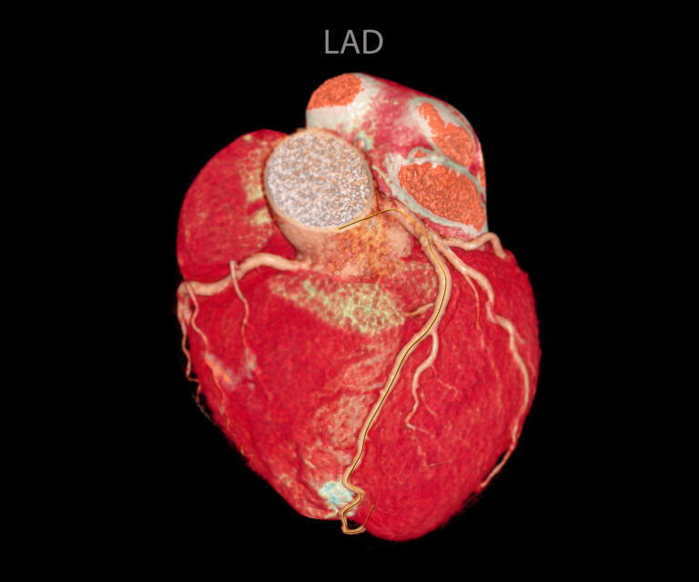
Angiography
Angiography uses contrast-enhanced X-ray imaging to visualise blood vessels and circulatory pathways. Also, it is used to support the diagnosis of vascular disease and guide interventional cardiac and peripheral procedures.
Mammography
Mammography applies low-dose X-ray imaging to examine breast tissue. This approach supports early cancer detection and evaluation of calcifications and structural changes.
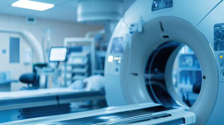
Computed Tomography
Computed Tomography (CT) combines X-ray acquisition with computer reconstruction to generate cross-sectional images. CT supports complex diagnosis, trauma assessment and detailed treatment planning.
Ultrasound
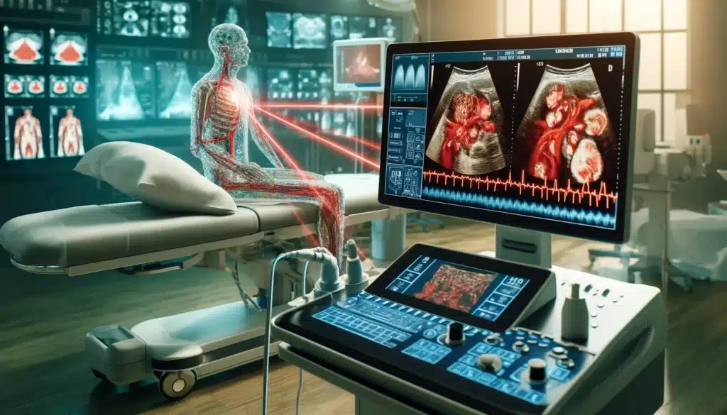
Ultrasound uses high-frequency sound waves to create real-time images of organs and soft tissues. This non-invasive technique is used in obstetrics, abdominal imaging and vascular assessment.
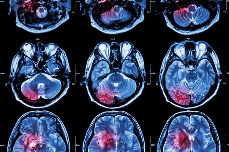
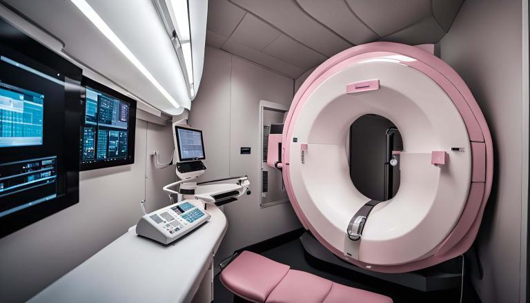
Magnetic Resonance Imaging
Magnetic Resonance Imaging (MRI) uses magnetic fields and radiofrequency signals to produce detailed soft-tissue images. MRI supports neurological, musculoskeletal and oncological evaluation without ionising radiation.
PET Imaging
Positron Emission Tomography (PET) uses radiotracers to measure metabolic and molecular activity within tissues. This imaging modality enables disease detection, staging and therapy monitoring in oncology, neurology and cardiology.
SPECT Imaging
Single Photon Emission Computed Tomography (SPECT) uses gamma-emitting radiotracers to assess physiological function. SPECT is applied in myocardial perfusion imaging and functional nuclear studies.
Hybrid Scanners
Hybrid scanners such as PET-CT and PET-MRI combine anatomical and functional imaging in a single examination. These scanners improve diagnostic confidence and enhance disease characterisation.
Fluoroscopy
Fluoroscopy provides continuous X-ray imaging to produce real-time moving images. This technique supports diagnostic studies and guides interventional procedures.
Tactile Imaging
Tactile Imaging converts pressure-sensor data into digital representations of soft-tissue structure. Also, it assists in the detection of abnormalities through mechanical property analysis.
Medical Photography
Medical Photography documents clinical conditions and procedures through high-resolution imaging. The aim is to support diagnosis, education and research activities.
Bone Densitometry
Bone Densitometry uses dual-energy X-ray absorptiometry to measure bone mineral density. This technique supports osteoporosis diagnosis and fracture risk assessment.
Optical Tomography
Optical Tomography produces high-resolution cross-sectional images of tissue microstructure. Also, it is used in ophthalmology for retinal assessment and disease monitoring.
Photoacoustic Imaging
Photoacoustic Imaging combines optical excitation with ultrasound detection to generate tissue images. This technique provides an insight into vascular and molecular features in research and emerging clinical applications.
EXPLORE
Structure and Function
Structural techniques, including X-ray, CT and MRI, provide detailed anatomical information. Ultrasound enables real-time assessment of organs and blood flow. PET and SPECT reveal metabolic and physiological activity beyond structure.
Hybrid platforms integrate anatomical and functional data into a single examination. This strengthens diagnostic confidence and disease characterisation. Artificial intelligence improves image quality, safety and accessibility.
CONTRIBUTE
Knowledge Gateway
The Imaging Modalities Hub provides access to expert-led features, innovation updates and clinical insights from Open MedScience. The hubs connects imaging disciplines, supporting informed decisions across research, education and practice.
Magnetic resonance imaging (MRI) revolutionized medical diagnostics by evolving from nuclear magnetic resonance discoveries to life-saving technology.
Understand the roles of medical scanners in modern medicine, highlighting their strengths and applications in healthcare diagnostics.
Magnetic Resonance Angiography Aorta provides clear insights into vascular diseases, enhancing treatment planning and follow-up care.
Mobile CT scanners are revolutionising healthcare by providing rapid, accessible diagnostics in remote, emergency, and disaster settings worldwide.
Uncover the latest innovations in photon-counting computed tomography and their impact on personalised healthcare and imaging.
Advances in medical imaging technology have significantly improved diagnostic accuracy, enabling earlier detection and more personalised treatments.
home » imaging modalities hub