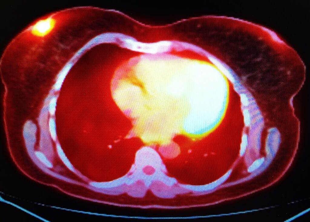
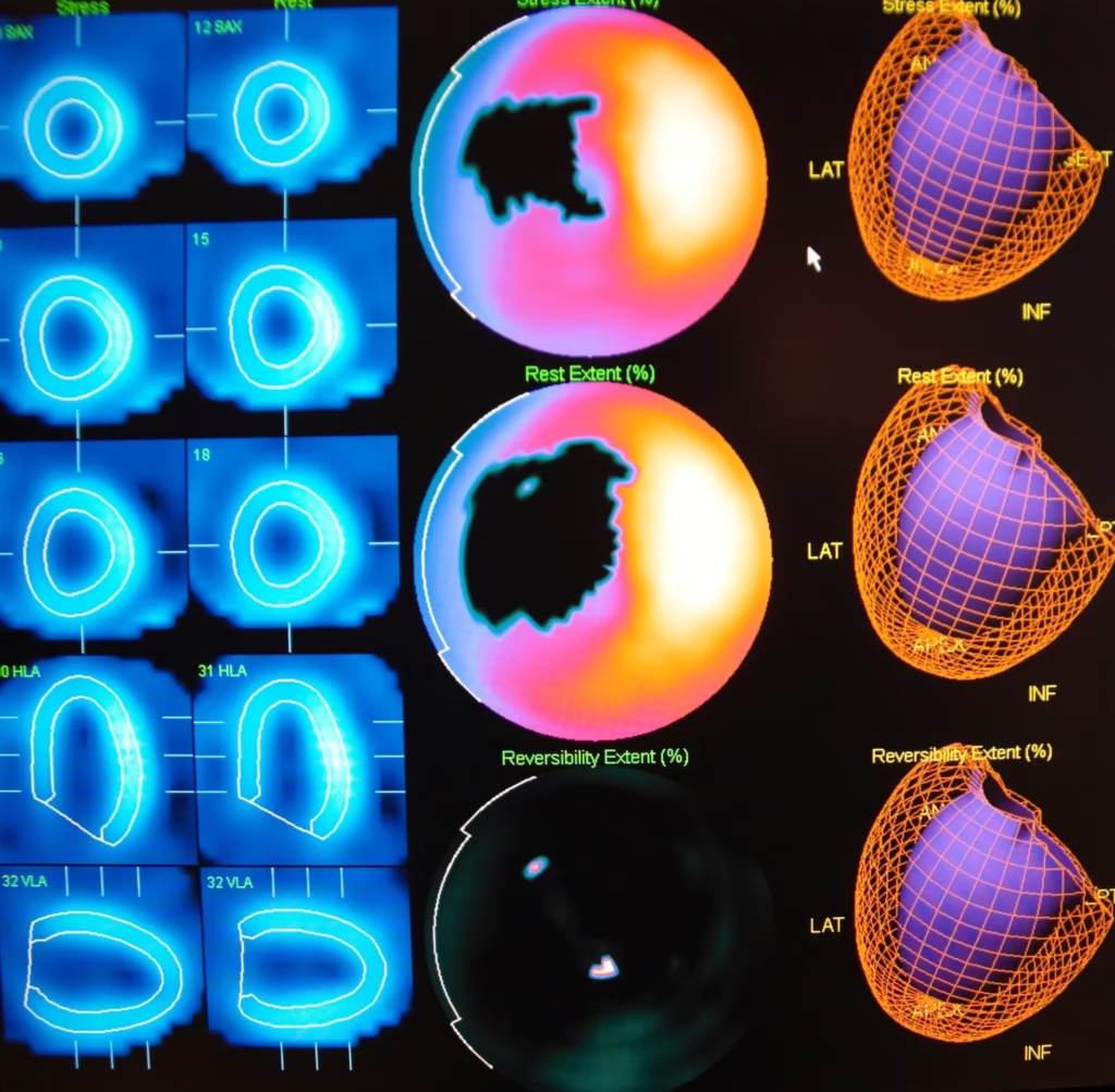
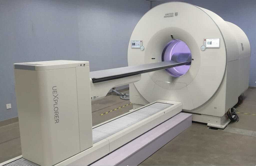
Nuclear medicine imaging is essential to detect and diagnose chronic disease, including cancer, but traditional methods like PET and SPECT aren’t perfect — they can’t always spot small details or abnormalities in the tissue. This is where bioengineered contrast agents, like nanoparticles, come in. These substances are designed or modified via biological engineering to improve image clarity and make nuclear medicine imaging more precise. Research has found that nanoparticles can help imaging agents stay in the bloodstream for up to 24 hours without being removed by the immune system. This means more of the contrast agent reaches the disease site, which results in sharper images. On top of that, AI can also play a role. Trained algorithms can quickly and accurately predict how various bioengineered contrast agents will act in the body, which helps researchers design even better ones.
Gold nanorods improve breast cancer imaging
Researchers in the U.S. recently bioengineered a nanoparticle that directly improves PET image contrast in breast cancer tumours in mice studies. To do this, they coated gold nanorods, a type of inorganic nanoparticle, with a mesoporous silica nanoshell coating. This coating has a large, porous surface somewhat similar to a sponge, which is what makes it so useful. As the nanoshell can hold a higher concentration of PET tracer, there’s a clearer signal for the PET scanner to pick up on, which results in clearer and brighter images.
To make them easier for the PET scanner to detect, the nanoparticles were then further labelled with a radionuclide (Zirconium-89). Radionuclides emit strong gamma rays, which PET scanners are highly attuned to. So, when even just a few of these radionuclides collect around a tumour or disease site, they make the area very visible. The researchers actually found that around 10% of the bioengineered contrast agent per gram accumulated in the breast cancer tumour. Although these promising findings are from mice studies, this contrast agent suggests great potential to help with future cancer diagnosis in humans.
AI in nuclear medicine imaging: the need for trust and accuracy
Artificial intelligence also holds a lot of promise to help advance the development of bioengineered contrast agents for nuclear medicine imaging. In fact, there’s been a recent surge of AI research in this field, with the number of AI-related papers published in radiology, nuclear medicine, and medical imaging now doubling every 2.7 years. But for AI to live up to its true potential, reliable and trustworthy systems are key. This is why the Society of Nuclear Medicine and Molecular Imaging have formed an AI task force — to guarantee that any AI-generated data is accurate and dependable. To achieve this, AI vendors should be transparent about how their systems are validated. This means they always need to provide clear details on the clinical tasks the algorithm has been trained on, the patient population they were tested on, and the specific protocols for image acquisition, reconstruction, and analysis. With this information, investors and nuclear medicine physicians can be confident they’re backing and using reliable AI tools.
Bioengineered contrast agents: AI-enhanced design
When AI tools are developed with accuracy and transparency in mind, the results are truly impressive. For example, AI can improve Monte Carlo simulations, which predict how bioengineered contrast agents will behave in the body, as well as how well the PET or SPECT scanner can detect them. Usually, Monte Carlo simulations are performed by computers, and this process takes a lot of time as all the possible random nanoparticle interactions are calculated. Fortunately, AI speeds up this process. Specifically, it can analyse vast volumes of Monte Carlo data to learn optimal pathways in the body for nanoparticles, and how well cells take them up. This, in turn, allows researchers to more easily identify the most effective nanoparticle designs to use for the clearest, most accurate images.
Still in early stages of development, bioengineered contrast agents are set to transform the future of precision in nuclear medicine imaging. These clever inventions make tumours and other diseases more visible for easier detection and diagnosis. And, with the help of AI, it’ll become even easier for researchers to develop agents that work as effectively and safely as possible.
Disclaimer
The content provided in this article is intended for informational and educational purposes only. It does not constitute medical advice, diagnosis, or treatment recommendations, and should not be used as a substitute for consultation with qualified healthcare professionals. The technologies and findings discussed, including the use of bioengineered contrast agents and artificial intelligence in nuclear medicine imaging, are based on ongoing research and are not necessarily approved for clinical use in humans. While promising results have been reported in preclinical studies, such as those involving animal models, further research and regulatory approval are required before these methods can be safely and effectively applied in clinical settings.
Open Medscience does not endorse or guarantee the safety, efficacy, or suitability of any specific product, method, or approach mentioned. Readers should exercise their own judgement and consult appropriate professionals before making decisions based on the information presented herein. Neither Open Medscience nor the authors shall be held liable for any damages or consequences resulting from the use of information provided in this article.
home » blog » nuclear medicine imaging »


