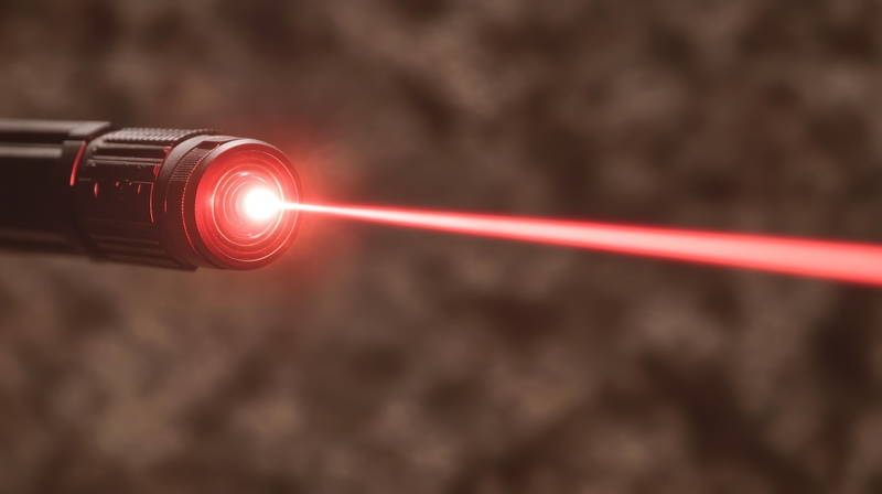
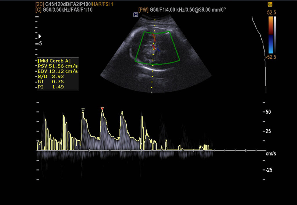
Medical lasers are used in various clinical applications, including cancer therapy and ophthalmology.
Beam Basics: The Science Behind How Lasers Operate
Max Planck showed that light is released in specific amounts of energy known as quanta and is related to radiation frequency (f). This discovery led to the equation E=hf, where h is Planck’s constant (6.6262 x 10 -34 Joule⋅second) and is fundamental to medical lasers.
Electromagnetic radiation is emitted from a charged particle (electron) that irradiates energy when it falls from a higher to a lower energy state. Its frequency or wavelength determines the colour of light. The shorter wavelengths are ultraviolet, and the longer wavelengths are infrared.
Quantum mechanics describes the smallest particle of light energy as a photon. The energy (E) of a photon is determined by its frequency (f) and Planck’s constant (h). The difference in energy levels across which an excited electron fall determines the wavelength of the emitted light.
The Planck equation set the foundation of the LASER (Light Amplification Stimulated Emission Radiation), and Albert Einstein 1916 described it as the theory of stimulated emission. This process involved an incoming photon of a specific frequency interacting with an excited atomic electron, causing it to relax back to a lower energy level. These fundamental discoveries of stimulated emission were the basis of the creation of modern lasers. However, it was not until 1960 that Maiman, a physicist at Bell Laboratories, generated a laser from ruby crystal (λ = 694 nm).
However, its practical capabilities began to emerge in the 1940s in the work of Charles Townes and Arthur Schawlow on the development of microwave spectroscopy.
This research led to the invention of the MASER (Microwave Amplification by Stimulated Emission of Radiation). First, however, Theodore Maiman created the first laser using an electrical source to energise a solid ruby.
More lasers were discovered, including the CO2 (carbon dioxide) laser, emitting a concentrated ray of light and absorbing water to vaporise tissue. In addition, the neodymium-yttrium aluminium garnet (Nd-YAG) laser-induced coagulative necrosis within the tissue. Also, visible light lasers were used to produce haemostasis.
Modern medical lasers are used in various clinical applications, including cancer therapy, ophthalmology, nerve stimulation, dermatology, plastic surgery, wound healing and dentistry.
However, the more advanced medical lasers based on the diode are used in cancer therapy. For example, these diode lasers are used in surgical procedures, including soft tissue cutting, coagulation and cancer thermal therapy.
Currently, different types of lasers are capable of undertaking invasive and non-invasive procedures at different depths. The type of laser depends on the region of the electromagnetic spectrum: UV (200-400) µm, visible (400-700) nm, near-IR (700-2900) nm and mid-IR (3-5) µm.
Since the conception of lasers, they have been classified according to the following characteristics:
- The laser medium where the amplification occurs can be a solid, liquid, or gas. These types of lasers are referred to as solid-state lasers, liquid lasers, or gas lasers.
- Gas lasers, including carbon dioxide, excimer (XeF) and argon, are used in medical lasers.
- Dye lasers are examples of liquid lasers, and ruby or yttrium aluminium garnet doped with neodymium lasers are solid-state lasers.
- Lasers can emit radiation at different wavelengths in the electromagnetic spectrum’s UV, visible, and IR parts. These are classified as UV lasers, visible lasers, and IR lasers.
- Lasers that emit radiation continuously are called continuous-wave (CW) lasers.
- Lasers that emit bursts of radiation are called pulsed lasers.
Lasers produce deeper tissue penetration depths in the near-infrared (750-1200) nm than visible lasers. Therefore, procedures that require deep penetrations, such as nano-gold mediated cancer therapy.
The visible lasers (430-680) nm have strong absorption in blood and have been used for oral cancer and retina decreases phototherapy.
Mid-IR lasers (1.9-3.0) µm and (9.3 -10.6) µm have strong absorption in water and tissue. This enables them to remove soft and hard tissue, a procedure known as ablation.
IR lasers (1.3-1.6) µm have been used for minimally invasive procedures such as resurfacing due to their more negligible tissue absorption.
Photoacoustic Imaging
Photoacoustic imaging is a hybrid imaging technique based on the photoacoustic effect. It delivers non-ionising laser pulses to tissues and produces heat which initiates thermoelastic expansion to generate an ultrasonic wave. These waves can be detected using ultrasound imaging equipment. This technique can be used to show organs and blood vessels to tumours. Also, more advanced photoacoustic imaging can picture 3-D images of internal body parts and differentiate cancerous cells from healthy cells. This technology platform is called Photoacoustic Topography Through an Ergodic Relay (PATER).
Clinical Applications of Medical Laser Technology
World’s Most Powerful Laser
The Thales and ELI-NP (Extreme Light Infrastructure for Nuclear Physics) projects have developed the world’s most powerful laser. The ultra-high-intensity laser system can produce a pulse at a peak power level of 10 petawatts (1015 W). This laser system is designed to generate twin laser beams of 10 PW and is used in nuclear physics. For example, this type of laser can be used to study key nuclear reactions relevant to nucleosynthesis. In particular, the fusion of α particles and carbon nuclei to produce oxygen (4He + 12C → 16O) is central to life on Earth.
The main features of the laser system are:
- The high-power laser system (HPLS) consists of two 10 PW beams that can deliver a laser pulse of up to 225 J in each arm during a laser pulse duration of 15–22.5 fs. The wavelength region is 814 – 825 nm.
- The maximum focal spot intensity is I0 ∼ 1023 W cm−2.
- The laser system can deliver lower powers at 100 TW and 1 PW.
- The Variable-Energy Gamma Ray (VEGA) system at ELI-NP can deliver monoenergetic gamma rays with up to 19.5 MeV.
- The primary performance parameters are a photon density of ∼104 s−1 eV−1, with a high degree of linear polarisation of >95%.
- The laser will be able to study the extreme states in gases, solids and plasma.
- The laser configuration can be altered to investigate systems that involve non-linear quantum electrodynamics (QED). In addition to vacuum birefringence, relativistically induced transparency (RIT) and nuclear resonance fluorescence (NRF). Including photoactivation, photonuclear reactions and photofission.
Laser Frontiers: The Convergence of Innovation in Medical, Industrial, and Defense Applications
The next generation of lasers will generate powerful beam sources at precisely the right wavelength for all medical and industrial applications. Since the laser conception, they are becoming smaller devices due to the advancement of semiconductors and direct diode lasers. This technology platform led to the Extreme Light Infrastructure for Nuclear Physics (ELI-NP) project, which uses the ultra-high intensity laser system to generate pulses at a peak power level of 10 petawatts. However, laser weapons based on electro-optical systems can increase the lethal power. For example, a high-energy laser system mounted on a US Army Boeing AH-64 Apache attack helicopter. Furthermore, the US Navy has developed solid-state lasers, including the Solid-State Laser Technology Maturation (SSL-TM) effort and High Energy Laser Counter-ASCM Program (HELCAP). However, continued research into LLLT on how to relate the dose to treat a range of clinical conditions in patients in a safe manner.
Disclaimer
The content presented in this article is intended for general informational and educational purposes only. While efforts have been made to ensure the accuracy of the scientific and historical information provided, Open Medscience makes no representations or warranties of any kind, express or implied, about the completeness, accuracy, reliability, suitability or availability with respect to the content or related graphics contained in the article for any purpose.
This article does not constitute medical advice, diagnosis, or treatment recommendations. Readers should not rely on the content as a substitute for professional medical advice or guidance from qualified healthcare professionals. Any medical or technical procedures mentioned should only be performed by trained and licensed professionals in appropriate clinical settings.
Open Medscience does not accept responsibility for any loss, damage, or injury that may result from the use of this information. The inclusion of any specific medical technologies, procedures, or historical accounts does not imply endorsement or recommendation.


