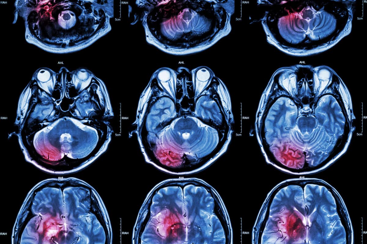
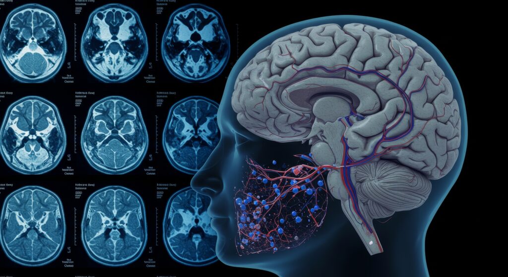
These non-radioactive labels can be incorporated into small molecules to study in vivo metabolic pathways in real-time.
Magnetic Resonance Imaging Of Cancer Metabolism
Hyperpolarization results from the nuclear spin polarization of the material when subjected to a magnetic field determined by the Boltzmann distribution. Hyperpolarization can make magnetic resonance’s signal-to-noise ratio (SNR) more prominent. Currently, hyperpolarization is being used in clinical studies to label drug candidates with carbon-13.
These non-radioactive labels can be incorporated into small molecules and can then be used to study in vivo metabolic pathways in real-time. For example, carbon-13 labelled pyruvate is becoming the gold standard when it comes to the development of hyperpolarized probes. Several preclinical models have demonstrated that hyperpolarized probes can be used in the areas of neurology and oncology. Therefore, these probes can be used in conjunction with magnetic resonance imaging (MRI) and consequently have great potential towards diagnosing a disease state in personalised medicine.
The output signals generated by MRI are weak compared to other imaging modalities, including imaging techniques based on acoustics, optics, and emission. However, several factors can be used to increase the SNR, especially by applying more powerful magnetic fields, high concentrations of spins, and longer acquisition times. MRI works on the human body because of its high concentration of water and, therefore, an abundance of protons. These protons have a relatively higher gyromagnetic ratio.
Hyperpolarization increases the signal response in magnetic resonance by increasing spin polarisation. Hyperpolarization aims to use the fundamental ability of nuclear magnetic resonance spectroscopy to identify chemical environments by chemical shift and characterise their dynamic properties in vivo.
NMR-Active Nucleus
The most abundant isotope of hydrogen is the proton, which has a spin of ½ a nucleus and therefore produces observable NMR signals. Consequently, the carbon-13 isotope of carbon possesses a spin of ½ a nucleus with an associated 1.1% natural abundance. To obtain a favourable NMR signal, it would be necessary to enrich the target molecule to increase SNR further isotopically.
The degree of hyperpolarization of the molecule will be the result of the location of the NMR-active nucleus. Therefore, the position of isotopic enrichment is a factor in the hyperpolarised molecule’s properties. For example, [1-13C] pyruvate molecules contain an enriched carbon-13 in position 1 of the molecule. Accordingly, this carbon-13 label would be the target of the hyperpolarization process through the nuclear spin.
The hyperpolarized carbon-13 atom will be transformed during the in vivo metabolic pathways, and each metabolite will produce a different signal. This approach offers great potential in ADME human studies because the hyperpolarized molecules can be used in vivo studies to determine the fate of the drug substance. The drug can be administered to the patient at physiological concentrations without harmful effects.
Novel Hyperpolarized Probes and Their Applications
In recent years, the field of hyperpolarized magnetic resonance imaging (MRI) has expanded significantly, with new probes being developed to target specific metabolic pathways and disease states. One such development is the use of hyperpolarized [2-13C] pyruvate, which offers a complementary metabolic pathway analysis to [1-13C] pyruvate by providing insights into different enzymatic steps of the tricarboxylic acid (TCA) cycle. This allows for a more comprehensive understanding of cellular metabolism, particularly in cancerous tissues.
Hyperpolarized [1-13C] α-ketoglutarate has also emerged as a promising probe, providing unique insights into mitochondrial metabolism and the redox state of cells. This probe is particularly useful in studying gliomas and other brain tumours, where metabolic rewiring is a hallmark of disease progression.
Advancements in Hyperpolarization Techniques
The field has seen significant advancements in the techniques used to achieve hyperpolarization. Dynamic Nuclear Polarization (DNP) remains the most widely used method. Still, newer techniques such as ParaHydrogen Induced Polarization (PHIP) and Signal Amplification By Reversible Exchange (SABRE) are gaining traction due to their ability to produce high polarization levels in a shorter time frame. These advancements are critical for making hyperpolarization more accessible for clinical applications.
Clinical Translation and Personalized Medicine
The transition from preclinical models to clinical applications is a critical step in the development of hyperpolarized MRI. The first human studies using hyperpolarized [1-13C] pyruvate have shown promising results, demonstrating the potential of this technology in diagnosing and monitoring treatment responses in prostate and breast cancers. The ability to non-invasively monitor metabolic changes in real-time offers a powerful tool for personalized medicine, allowing for tailored treatment plans based on the metabolic profile of an individual’s tumour.
Regulatory and Safety Considerations
As with any new medical technology, regulatory and safety considerations are paramount. The non-radioactive nature of hyperpolarized probes is a significant advantage, reducing the risk associated with traditional radiolabeled compounds. However, ensuring the safety and efficacy of these probes in humans requires rigorous clinical trials. The metabolic fate of hyperpolarized compounds must be thoroughly understood to avoid potential toxicity or unintended biological effects.
Future Directions
The future of hyperpolarized MRI lies in developing new probes and refining existing ones to target a broader range of metabolic pathways. Research is currently underway to develop hyperpolarized probes for amino acids, lipids, and other key metabolic intermediates. These new probes could provide insights into diseases beyond cancer, such as neurological disorders, cardiovascular diseases, and metabolic syndromes.
Moreover, combining hyperpolarized MRI with other imaging modalities, such as positron emission tomography (PET) or computed tomography (CT), could offer a more comprehensive picture of disease states, leveraging the strengths of each technique. The integration of artificial intelligence (AI) and machine learning algorithms for data analysis could further enhance the diagnostic and prognostic capabilities of hyperpolarized MRI.
Conclusion
The imaging properties of hyperpolarized molecules are a function of their relaxation properties and ease of hyperpolarization. Other factors include the safety profile, biological availability, and metabolism profile. Several molecules, including carbon-13 labelled pyruvate, have been subjected to hyperpolarization, and their associated metabolism was imaged. These hyperpolarized probes include [1,4-13C2]fumarate used to evaluate cell necrosis and [U-2H, U-13C]glucose to assess the glycolytic and pentose phosphate pathway activities and detect early treatment response. In addition, 13C-labelled bicarbonate was used for in vivo pH mapping, including 13C-labelled urea as a marker of perfusion.
Disclaimer
The content presented in this article is intended for informational and educational purposes only. It does not constitute medical advice, diagnosis, or treatment recommendations. The technologies and procedures described, including the use of hyperpolarized carbon-13 labelled compounds for magnetic resonance imaging, are subject to ongoing research and clinical validation. Any mention of diagnostic or therapeutic applications is not a substitute for professional medical consultation or regulatory approval. Readers are advised to consult qualified healthcare professionals or appropriate regulatory authorities before relying on the information provided herein. Open Medscience does not assume responsibility for the accuracy, completeness, or current relevance of the content, particularly where clinical use is concerned.


