
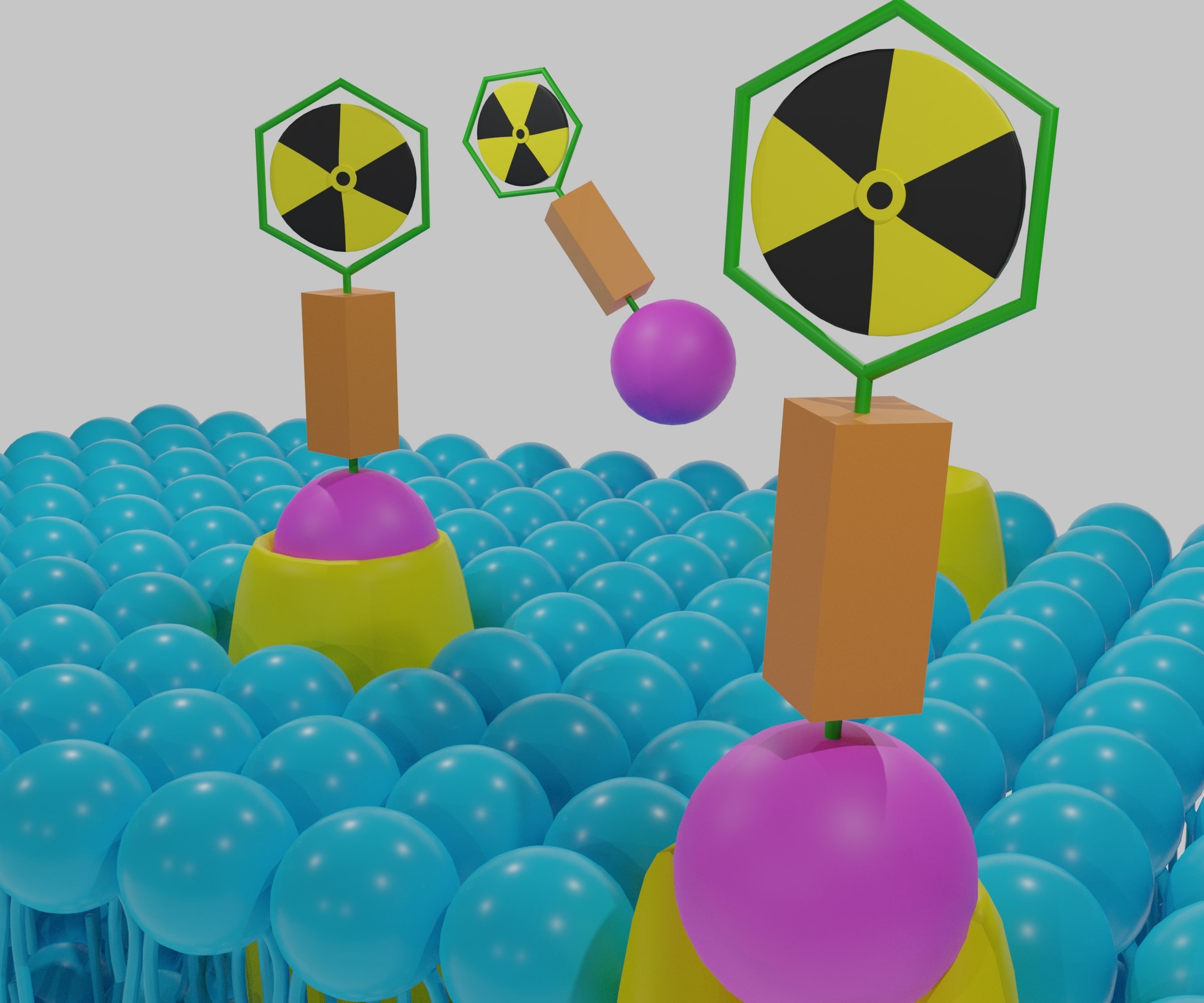
Radiotheranostics: The Dual Power of Diagnosis and Therapy in Modern Medicine
 By
By
Learn how radiotheranostics in cancer care combines imaging and therapy for better outcomes in advanced malignancies.
By
Learn how radiotheranostics in cancer care combines imaging and therapy for better outcomes in advanced malignancies.
 By
By
Radiotheranostics and radiotherapeutics cancer treatments provide personalised care by combining diagnosis and targeted therapy.
 By
By
Yttrium-90 OPS201, a receptor antagonist, delivers targeted beta radiation for effective neuroendocrine tumour therapy and research.
 By
By
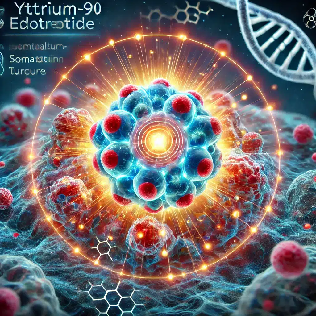
Yttrium-90 Edotreotide delivers targeted beta radiation therapy to somatostatin receptor-positive neuroendocrine tumours with precision.
 By
By
Yttrium-90 DOTALAN delivers targeted beta radiation to somatostatin receptor-expressing tumours, treating neuroendocrine neoplasms effectively.
 By
By
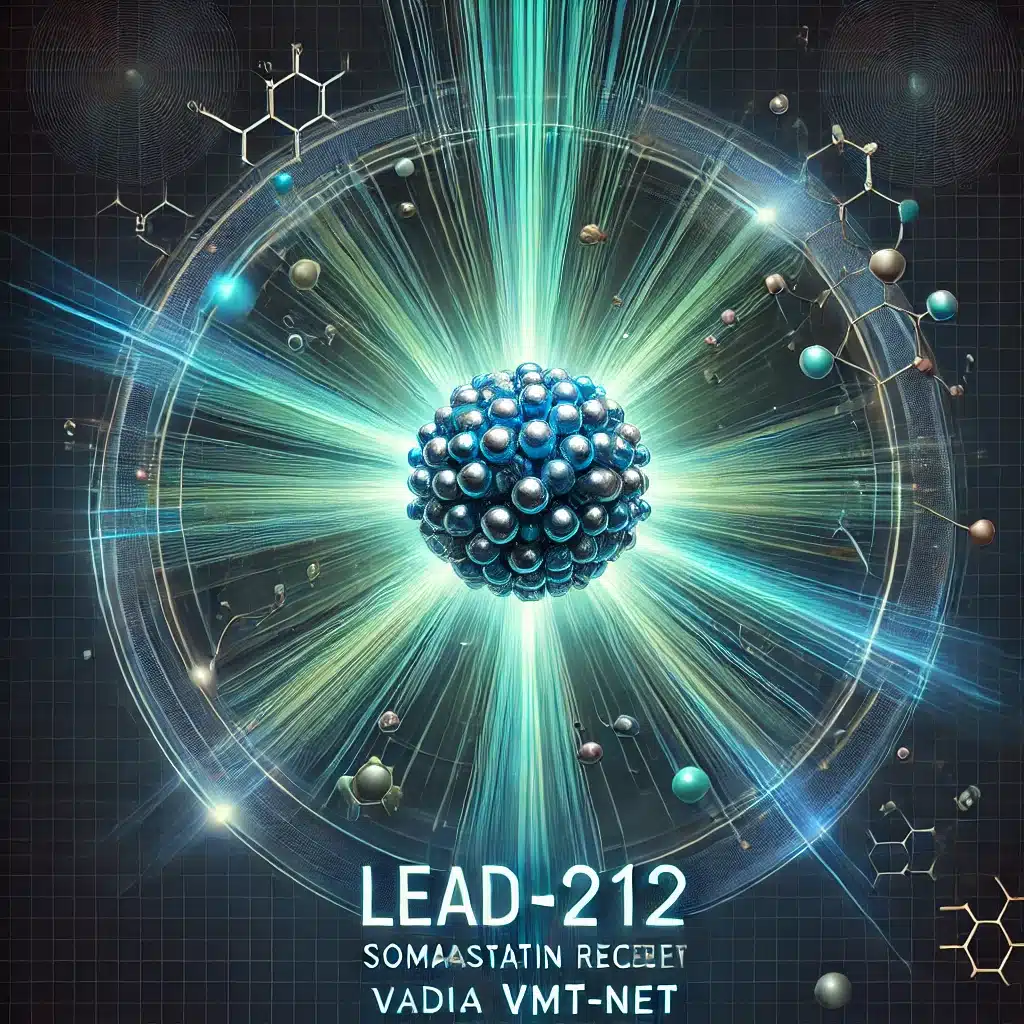
Lead-212 VMT-α-NET offers advanced targeted therapy for neuroendocrine tumours by harnessing precise alpha-particle radiation.
 By
By
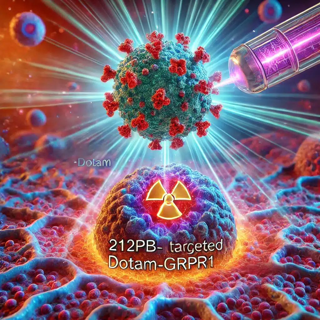
Lead-212 DOTAM-GRPR1 combines targeted GRPR binding with alpha radiation, offering innovative treatment for resistant cancers.
 By
By
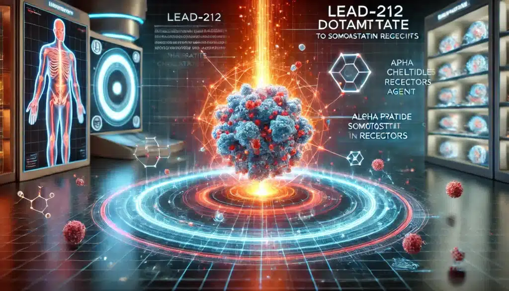
Lead-212 DOTAMTATE targets somatostatin receptors in neuroendocrine tumours, delivering potent alpha-emission therapy with minimal off-target toxicity.
 By
By
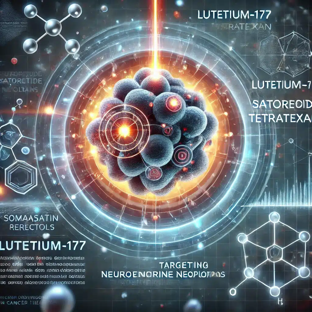
Lutetium-177 Satoreotide Tetratexan is a radiopharmaceutical targeting somatostatin receptors, providing advanced therapeutic options for neuroendocrine neoplasms effectively.
 By
By
Lutetium-177 Ludotadipep targets PSMA with enhanced stability, specificity, and reduced side-effects, revolutionising prostate cancer treatment.
 By
By
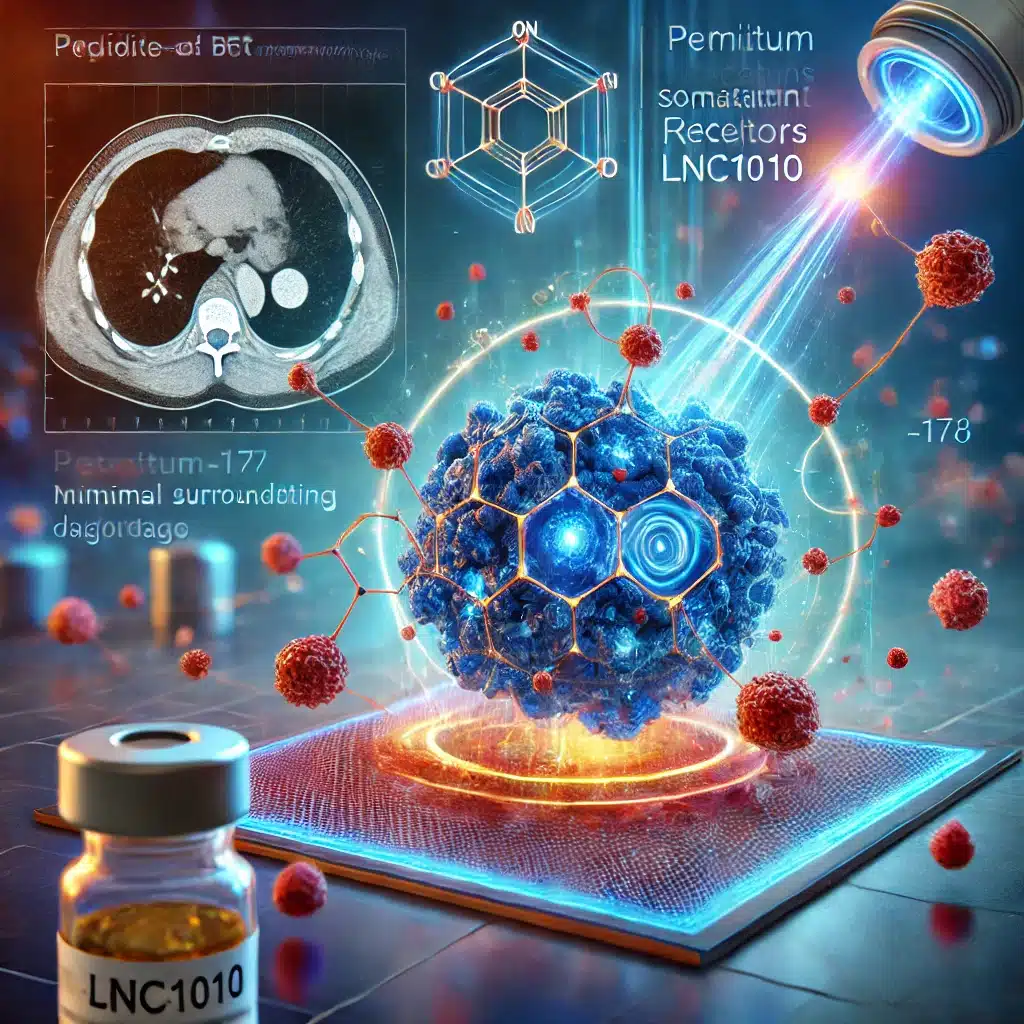
Lutetium-177 LNC1010, a radiolabelled therapy, targets somatostatin receptors, delivering precise treatment with minimal side effects.
 By
By
Lutetium-177 Edotreotide revolutionises neuroendocrine tumour treatment through targeted radioligand therapy, enhancing precision and improving patient outcomes.
 By
By
Lutetium-177 DTPA-Omburtamab offers hope in neuroblastoma treatment by precisely targeting B7-H3, delivering localised radiation while sparing healthy tissues.
 By
By
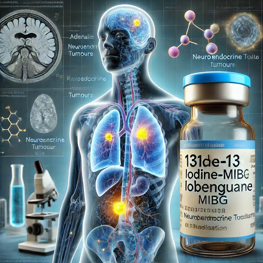
Iodine-131 Iobenguane revolutionises neuroendocrine tumour management by offering targeted imaging and therapy, significantly improving diagnosis, treatment, and patient outcomes.
 By
By
The REAL-LU study highlights Lutetium-177 DOTATATE’s real-world effectiveness, safety, and quality-of-life impact in Italian patients with GEP-NETs.
 By
By
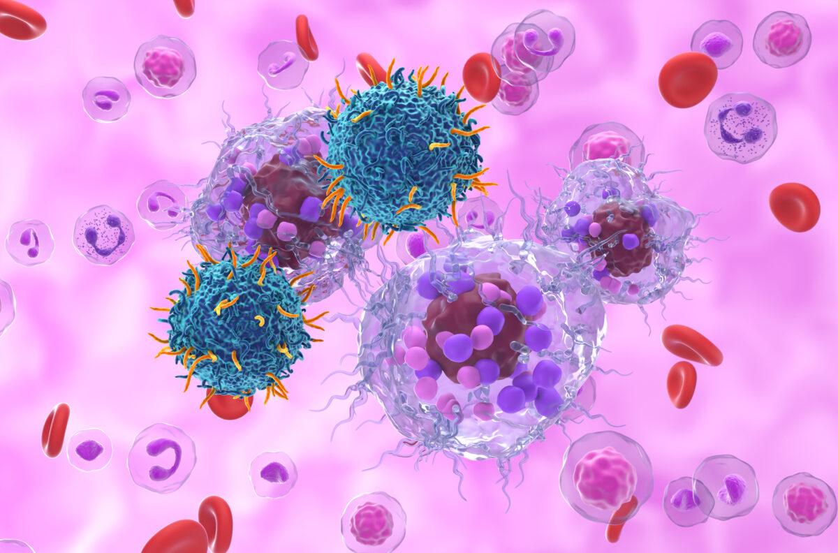
Neuroblastoma Targeting Agents offer innovative diagnostic and therapeutic options, revolutionizing treatment with precise targeting and efficacy.
 By
By
Somatostatin Receptor Targeting Agents offer groundbreaking diagnostic and therapeutic solutions for neuroendocrine tumour management and care.
 By
By
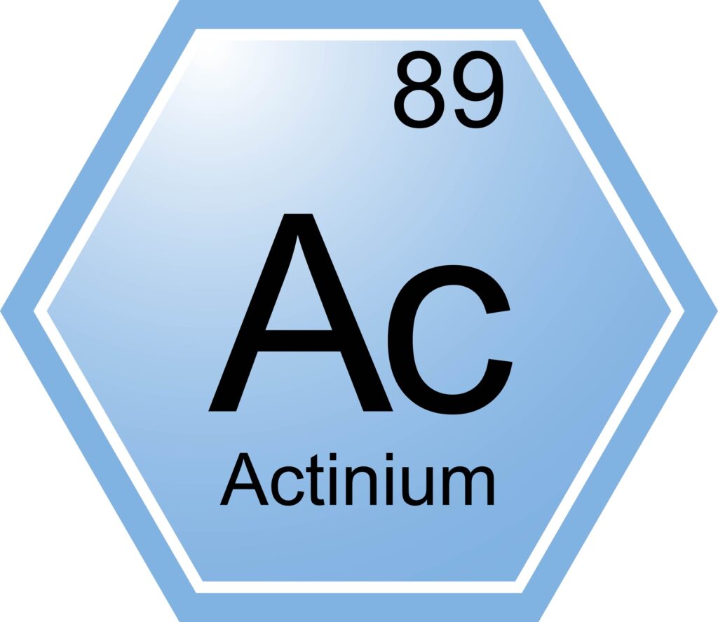
225Ac-RYZ101 offers promising GEP-NET treatment, targeting tumours with alpha particles via somatostatin receptor-specific edotreotate.
 By
By
Gallium-68 DOTATATE, a radiopharmaceutical, targets somatostatin receptors, aiding neuroendocrine tumor detection through PET imaging, enhancing diagnostic accuracy and patient outcomes.

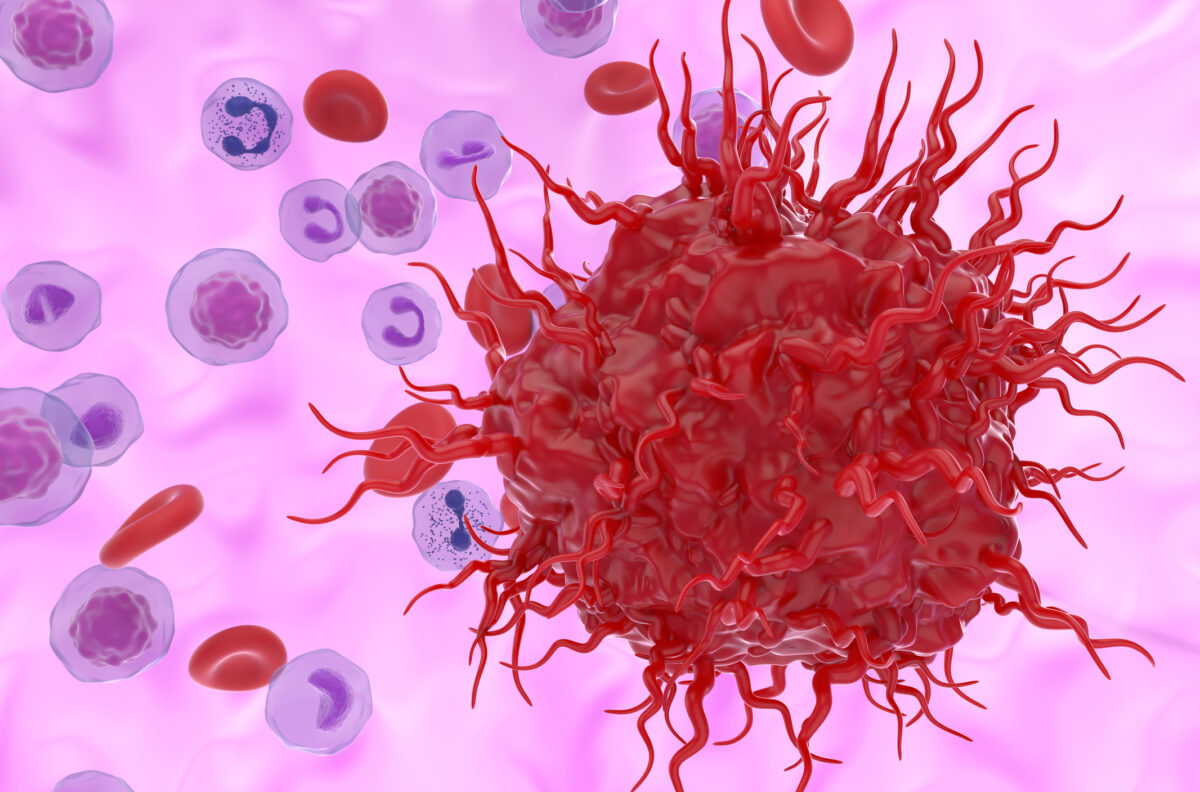
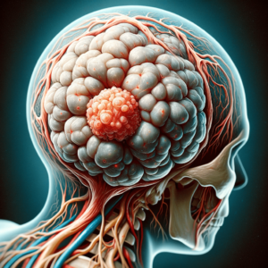
Neuroendocrine tumours include a spectrum of neoplasms characterized by histologic heterogeneity.