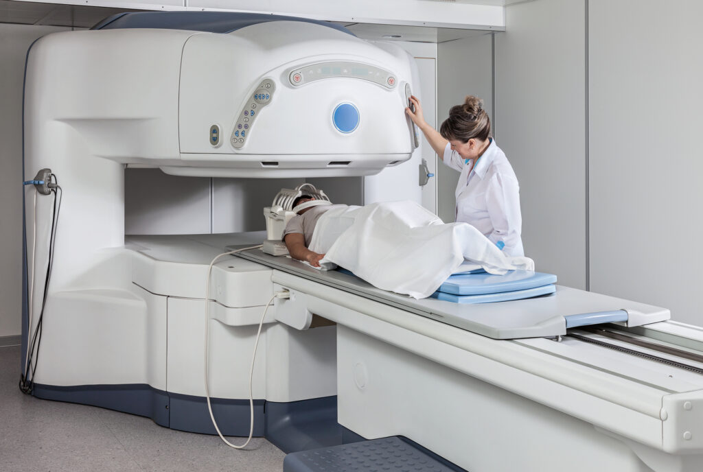
Radiopharmaceuticals in medical imaging combine radionuclides with pharmaceuticals for targeted diagnostic imaging and therapy.
Introduction to Radiopharmaceuticals
Radiopharmaceuticals, a unique category of drugs, have been pivotal in revolutionising the field of medical imaging. These substances, which are composed of a radioactive component and a pharmaceutical component, are designed to target specific organs, tissues, or cellular receptors in the body. The significance of radiopharmaceuticals lies in their ability to provide crucial information about the physiology and pathology of various systems within the body, thereby aiding in accurate diagnosis, staging, and treatment monitoring of numerous diseases.
Radiopharmaceuticals, a cornerstone in the field of nuclear medicine, are composed of two primary components: a radionuclide and a pharmaceutical agent. The radionuclide, a radioactive isotope, is the key element that enables imaging. It emits radiation that is detectable by specialised medical imaging equipment, such as PET or SPECT scanners. This radiation forms the basis for creating visual representations of internal biological processes. The pharmaceutical component of radiopharmaceuticals plays a crucial role in guiding the radionuclide to specific cells or tissues. It ensures that the radioactivity is targeted to the area of interest, thereby providing specific, localised images or therapeutic effects.
Radionuclides in radiopharmaceuticals are chosen based on their radiation characteristics, half-life, and the specific diagnostic or therapeutic application. The radiation emitted can be alpha particles, beta particles, or gamma rays, each with unique properties and uses.
- Technetium-99m: Widely used due to its ideal half-life (about 6 hours) and gamma emission, which is suitable for diagnostic imaging. It’s used in a variety of scans, including bone scans, heart scans, and brain imaging.
- Fluorine-18: Primarily used in PET scans, particularly in the form of fluorodeoxyglucose (FDG), which is valuable in cancer imaging due to its role in highlighting glucose metabolism.
- Iodine-131: Known for its beta emissions and longer half-life, it’s used both for diagnostic imaging and treatment, especially in thyroid conditions.
The localisation of radiopharmaceuticals within the body is governed by various mechanisms, ensuring that they accumulate in the target tissue or organ.
- Active Transport: Certain radiopharmaceuticals are actively transported into cells, often exploiting the natural cellular uptake mechanisms, like glucose or amino acids.
- Passive Diffusion: Some compounds passively diffuse into tissues, particularly in areas with increased permeability, such as inflamed or cancerous tissues.
- Receptor Binding: Specific radiopharmaceuticals are designed to bind to particular cellular receptors, enabling targeted imaging or therapy of cells that express these receptors.
- Phagocytosis: In some cases, especially in infection imaging, radiopharmaceuticals are taken up by phagocytes, cells that engulf foreign particles.
The effectiveness of localisation and the quality of the resulting images or therapeutic effect depend significantly on the biological characteristics of the target tissue and the properties of the radiopharmaceutical used. This precision in targeting allows for highly detailed and functional imaging, making radiopharmaceuticals invaluable in modern medicine for diagnosis, staging, and treatment monitoring across a wide range of diseases.
Imaging Techniques Using Radiopharmaceuticals
Positron Emission Tomography (PET)
Positron Emission Tomography, commonly known as PET, is a highly sophisticated imaging technique that plays a crucial role in various medical fields, including oncology, neurology, and cardiology. One of the most common radiopharmaceuticals used in PET imaging is the Fluorine-18 labelled glucose analogue, FDG (Fluorodeoxyglucose). FDG is a glucose analogue with a normal hydroxyl group replaced with 18F, a radioactive fluorine isotope.
- Oncology PET imaging is instrumental in detecting and staging cancerous tumours. Cancer cells, known for their high metabolic rate, absorb more glucose than normal cells. FDG accumulates in these high-metabolism areas, making locating and assessing tumours easier. PET scans are also critical in evaluating treatment response and monitoring recurrence.
- Neurology and PET imaging provide valuable insights into brain disorders. It helps in the diagnosis and management of conditions like Alzheimer’s disease, Parkinson’s disease, and epilepsy by assessing brain metabolism and function.
- Cardiology and PET scans are used to evaluate myocardial perfusion. They can identify areas of reduced blood flow to the heart, aiding in diagnosing and managing coronary artery disease and other cardiac conditions.
Single Photon Emission Computed Tomography (SPECT)
SPECT imaging is another vital nuclear medicine technique that utilises gamma rays for imaging. SPECT imaging commonly uses Technetium-99m labelled compounds and provides valuable diagnostic information in various specialties.
- In cardiology, SPECT is widely used to assess cardiac perfusion. It helps identify areas of the heart muscle that are not receiving adequate blood supply, which is crucial in diagnosing and managing coronary artery diseases.
- In neurology, SPECT imaging assists in evaluating brain blood flow. It is particularly useful in the assessment of stroke, dementia, and epilepsy, providing critical information about cerebral perfusion and brain function.
SPECT is also extensively used in orthopaedics and oncology for bone imaging. It is especially effective in detecting bone metastases, as cancerous cells in bone tend to absorb more of the radiopharmaceutical, revealing their presence in the SPECT images.
Beyond PET and SPECT, there are other notable imaging techniques involving radiopharmaceuticals.
- Gamma Imaging: This involves the use of gamma cameras to detect gamma radiation emitted by radiopharmaceuticals. It’s used for a variety of purposes, including thyroid, lung, and kidney imaging.
- Theranostics: This emerging field combines diagnostic imaging and therapeutic treatment. It involves using radiopharmaceuticals that not only image but also treat diseases. A prime example is the use of radioactive iodine in the treatment of thyroid disorders, where it is used both for imaging thyroid function and destroying thyroid cancer or overactive thyroid cells.
These techniques showcase radiopharmaceuticals’ versatility and efficacy in diagnosing and treating a wide range of diseases, highlighting their indispensable role in modern medicine.
Clinical Applications
Radiopharmaceuticals, which are unique medicinal formulations containing radioisotopes, are of great significance in modern medicine, particularly in diagnostics. Let’s explore their applications in various fields:
- Oncology: In cancer care, radiopharmaceuticals are indispensable. PET (Positron Emission Tomography) imaging using FDG (Fluorodeoxyglucose) is a standard procedure for evaluating various cancers, including lymphoma, lung cancer, and breast cancer. This technique helps in accurately detecting cancer, determining its stage, and monitoring the effectiveness of treatments.
- Cardiology: In the field of heart health, SPECT (Single Photon Emission Computed Tomography) and PET imaging play vital roles. They are primarily used in diagnosing coronary artery disease, evaluating myocardial infarction (heart attacks), and assessing the viability of heart muscle tissue. These imaging techniques provide crucial information about the health and function of the heart.
- Neurology: Radiopharmaceuticals are also essential in diagnosing and studying neurological conditions. They are used in identifying and assessing diseases like Alzheimer’s, Parkinson’s, epilepsy, and brain tumours. This is particularly important for early detection and treatment planning.
- Other Applications: Beyond these key areas, radiopharmaceuticals have a variety of other applications. They are used in thyroid imaging, which can help in diagnosing and managing thyroid diseases. In renal function assessment, they assist in evaluating how well the kidneys are working. Additionally, they are useful in localising infections and inflammations within the body.
Radiopharmaceuticals offer a non-invasive yet highly effective means to diagnose and monitor a wide range of health conditions, making them a crucial tool in modern medical practice.
Safety and Regulatory Aspects
The use of radiopharmaceuticals, while offering significant benefits in diagnostics and treatment, also involves certain safety considerations and is subject to a strict regulatory framework:
Safety Considerations:
- Radiation Exposure: One of the primary concerns with radiopharmaceuticals is the risk associated with radiation exposure. This risk, however, is generally low and is carefully managed.
- Selection of Radionuclides: The key to minimising radiation risk lies in the careful selection of radionuclides. These are chosen based on their half-lives and energy levels, which are optimised to deliver effective diagnostic or therapeutic results while minimising radiation exposure to the patient.
- Adverse Reactions: While adverse reactions to radiopharmaceuticals are possible, they are relatively rare and usually mild when they do occur. The low incidence of adverse reactions is attributed to these compounds’ meticulous development and testing.
Regulatory Framework
- Development and Production Oversight: The development and production of radiopharmaceuticals are rigorously regulated. Regulatory bodies like the U.S. Food and Drug Administration (FDA) and the European Medicines Agency (EMA) play pivotal roles in this oversight.
- Safety, Efficacy, and Quality Assurance: The regulations set by these bodies are designed to ensure the safety, efficacy, and quality of radiopharmaceuticals. This involves stringent testing and approval processes before these substances can be used clinically.
- Continual Monitoring: Besides the initial approval, ongoing monitoring ensures that any long-term effects or unforeseen issues are identified and addressed.
The combination of safety considerations and a robust regulatory framework ensures that radiopharmaceuticals, while inherently involving radioactive substances, are used to maximise patient benefit while minimising risks. This careful balance is key to the successful integration of these advanced medicinal products into modern medical practices.
Future Directions and Innovations
The field of radiopharmaceuticals is witnessing significant advances and developments, promising to revolutionise the way diseases are diagnosed and treated:
Advances in Radiopharmaceuticals
- Improved Targeting Capabilities: Researchers are focusing on developing new radiopharmaceuticals that can more accurately target specific cells or tissues. This specificity can lead to more effective diagnosis and treatment of diseases, particularly in oncology.
- Reduced Radiation Exposure: A key development area is reducing the radiation dose required for effective imaging or treatment. This involves creating radiopharmaceuticals with shorter half-lives or lower radiation levels, minimising the exposure for patients.
- Enhanced Imaging Quality: Enhanced image quality allows for more precise and detailed visualisation of diseases. This is crucial for accurately diagnosing, staging, and monitoring various medical conditions.
Emerging Applications:
- Personalised Medicine: One of the most exciting applications of radiopharmaceuticals is in personalised medicine. This approach involves using radiopharmaceuticals to tailor treatment plans based on the unique characteristics of each patient, such as their specific type of cancer or the genetic makeup of their disease.
- Radiopharmaceuticals in Immunotherapy: Integrating radiopharmaceuticals with immunotherapy is an emerging field. This approach aims to boost the effectiveness of immunotherapy by using radiopharmaceuticals to either directly target cancer cells or modulate the immune system.
Technological Developments
The advancements in imaging technology, particularly in the field of PET scanners and hybrid imaging systems, are playing a transformative role in enhancing the effectiveness of radiopharmaceuticals in medical diagnostics:
High-Resolution PET Scanners
- Resolution and Speed Improvements: The latest PET scanners boast significantly higher resolution and faster imaging capabilities. This means that they can provide much clearer and more detailed images, which is crucial for accurately detecting and characterising diseases at earlier stages.
- Diagnostic Accuracy: These PET technology improvements enhance radiopharmaceuticals’ diagnostic accuracy. Higher resolution images allow for a more precise visualisation of how radiopharmaceuticals accumulate in various tissues, leading to better identification and assessment of diseases.
Hybrid Imaging Systems
- Combining Imaging Modalities: Hybrid imaging systems merge the capabilities of PET or SPECT (Single Photon Emission Computed Tomography) with other imaging techniques, such as CT or MRI. Each of these modalities has its strengths; for instance, PET and SPECT excel in showing functional and metabolic processes, while CT and MRI are better at depicting anatomical structures.
- Comprehensive View of the Body: By combining these technologies, hybrid systems provide a more comprehensive view of the body’s internal workings. This integration is particularly beneficial for complex diseases like cancer, where understanding both the anatomical and functional aspects is crucial for accurate diagnosis and treatment planning.
- Improved Localisation and Characterisation: The combination of functional and anatomical imaging allows for better localisation and characterisation of disease processes. For example, a hybrid PET/CT scan can show not only the metabolic activity of a tumour (using PET) but also its exact size and location (using CT).
These technological advancements significantly enhance the utility of radiopharmaceuticals in medical diagnostics. They enable more accurate disease detection, staging, and monitoring, which are essential for effective treatment and management of various health conditions.
These advancements in radiopharmaceuticals and related technologies signify a significant leap forward in medical diagnostics and therapeutics. They offer the potential for more accurate diagnoses, more effective treatments, and the promise of highly personalised medical care.
Conclusion
Radiopharmaceuticals have emerged as an essential component in the area of medical imaging, offering unparalleled insights into a myriad of physiological and pathological processes. These specialised compounds, marked by their inclusion of radioactive isotopes, have revolutionised diagnostic methodologies, allowing healthcare professionals to visualise and analyse bodily functions and abnormalities with remarkable precision.
The fundamental principle behind radiopharmaceuticals lies in their ability to target specific cells or tissues within the body. Once administered, these compounds emit radiation that can be detected and visualised using imaging technologies such as Positron Emission Tomography (PET) and Single Photon Emission Computed Tomography (SPECT). This approach is particularly effective in identifying and assessing cancerous growths, cardiac anomalies, neurological disorders, and other internal pathologies.
What sets radiopharmaceuticals apart is their dual functionality. Not only do they enhance the visualisation of diseases, but they also provide crucial functional information. For instance, in oncology, they can indicate the metabolic activity of tumours, thus offering insights into the aggressiveness of cancer and the effectiveness of treatments. In cardiology, they assist in evaluating blood flow to the heart muscle, aiding in diagnosing coronary artery disease and assessing damage post-myocardial infarction.
The integration of radiopharmaceuticals with advanced imaging technologies like high-resolution PET scanners and hybrid imaging systems, such as PET/CT and PET/MRI, has further expanded their diagnostic capabilities. These technological advancements provide a more comprehensive view of the body by combining anatomical and functional imaging, thus enhancing the accuracy of diagnoses and the precision in treatment planning.
Looking to the future, the continued development of radiopharmaceuticals holds immense promise. Emerging trends include the creation of novel compounds with better-targeting abilities, reduced radiation exposure, and the potential for personalised medicine. These advancements could lead to more accurate early detection of diseases, tailored treatment regimens, and improved monitoring of treatment response.
Radiopharmaceuticals represent a transformative force in medical imaging. Their ability to provide detailed and functional insights into various bodily processes, coupled with ongoing advancements in imaging technology, positions them as a pivotal tool in enhancing diagnostic accuracy and patient care. As research and development in this field continue to advance, the potential benefits for patient outcomes and healthcare systems worldwide are both significant and exciting.
Disclaimer
The content provided in this article, The Transformative Impact of Radiopharmaceuticals in Medical Imaging, is intended for informational and educational purposes only. It does not constitute medical, scientific, or professional advice and should not be relied upon as such. While efforts have been made to ensure accuracy at the time of publication (12 January 2024), Open Medscience makes no warranties regarding the completeness, reliability, or applicability of the information presented.
Radiopharmaceuticals involve regulated substances and procedures that should only be administered or interpreted by qualified healthcare professionals in appropriate clinical settings. Readers should not attempt to use or apply any of the described techniques or substances without professional guidance and oversight.
Open Medscience disclaims any liability for loss, damage, or injury incurred as a result of the use of, or reliance on, information provided in this article. For specific medical advice, diagnoses, or treatment, please consult a licensed healthcare provider.
home » blog » medical imaging topics ».


