Radiopharmaceuticals are specialised compounds used in the field of nuclear medicine for diagnostic imaging and therapy. This article looks into the world of radiopharmaceuticals, focusing on their diagnostic applications. It begins by introducing the fundamental concepts of radiopharmaceuticals and the science of nuclear medicine. The article then explores the various types of radiopharmaceuticals used for diagnostics, the mechanisms by which they operate, and their clinical applications in different imaging modalities. The safety, production, and regulatory aspects of radiopharmaceuticals are also discussed, along with emerging trends and future directions in the field. The article concludes by reflecting on the potential advancements in radiopharmaceuticals and their significance in personalised medicine.
Introduction to Radiopharmaceuticals
Radiopharmaceuticals are a unique class of drugs that contain radioactive isotopes and are used primarily in the field of nuclear medicine. These compounds have a pivotal role in both diagnostic imaging and therapeutic applications. By emitting radiation, they enable the visualisation of physiological processes within the body, facilitating the diagnosis of various conditions and diseases.
The Science of Nuclear Medicine
Nuclear medicine is a medical specialty that uses small amounts of radioactive substances, known as radiotracers, to diagnose and treat diseases. This branch of medicine combines chemistry, physics, mathematics, computer technology, and medicine. Unlike conventional imaging techniques, such as X-rays or MRI, which provide structural information, nuclear medicine offers insights into the functional aspects of organs and tissues.
Types of Radiopharmaceuticals for Diagnostics
Radiopharmaceuticals used for diagnostic purposes are designed to target specific organs, tissues, or cellular receptors, depending on the clinical question being addressed. The primary types of radiopharmaceuticals can be categorised based on the type of radiation they emit and their application in various imaging modalities.
Gamma Emitters
Gamma-emitting radiopharmaceuticals are the most commonly used in nuclear medicine diagnostics. These compounds emit gamma rays, which can be detected by imaging devices such as gamma cameras and single-photon emission computed tomography (SPECT) scanners. Technetium-99m (Tc-99m) is the most widely used gamma emitter, valued for its ideal half-life and energy properties. Tc-99m is employed in various imaging procedures, including bone scans, cardiac imaging, and the detection of infections and tumours.
Positron Emitters
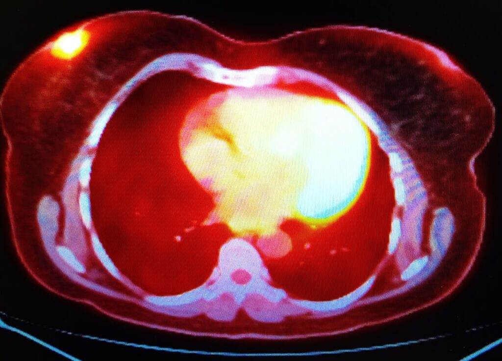
Positron-emitting radiopharmaceuticals are utilised in positron emission tomography (PET) imaging. These compounds emit positrons, which interact with electrons in the body, leading to the production of gamma rays that are detected by the PET scanner. Fluorine-18 (F-18) is one of the most commonly used positron emitters, with Fluorodeoxyglucose (FDG) being a notable example. FDG-PET imaging is widely used in oncology, neurology, and cardiology to assess metabolic activity and detect malignancies, brain disorders, and heart conditions.
Radiopharmaceuticals for Hybrid Imaging
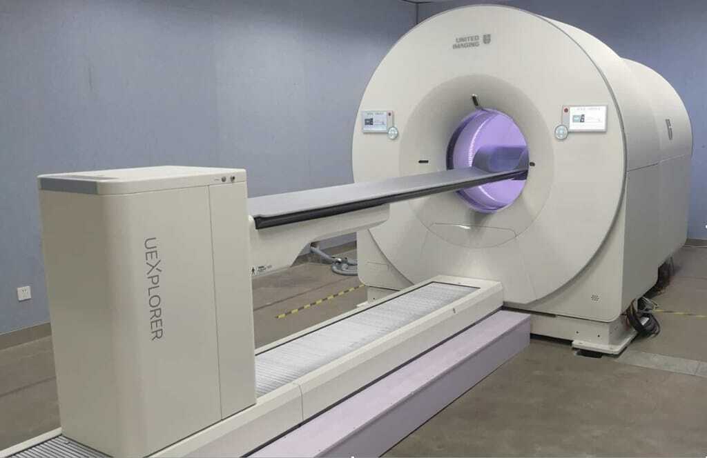
Hybrid imaging modalities, such as PET/CT and SPECT/CT, combine functional imaging with anatomical imaging. These modalities utilise radiopharmaceuticals that can be visualised by both PET or SPECT and computed tomography (CT) scanners. The integration of functional and anatomical data enhances the accuracy of diagnosis and allows for better localisation of abnormalities.
Mechanisms of Radiopharmaceuticals in Diagnostics
The diagnostic capability of radiopharmaceuticals is based on their ability to mimic physiological processes or to bind to specific cellular targets. The following mechanisms outline how these compounds function in the body to provide diagnostic information.
Physiological Distribution
Some radiopharmaceuticals are distributed throughout the body based on their physiological properties. For example, FDG, a glucose analogue, accumulates in cells with high glucose metabolism, such as cancer cells. By detecting the areas of high FDG uptake, PET imaging can reveal the presence of tumours or regions of abnormal metabolic activity.
Receptor Binding
Certain radiopharmaceuticals are designed to bind specifically to cellular receptors. For instance, radiolabelled peptides that target somatostatin receptors are used to image neuroendocrine tumours. The specific binding of these radiopharmaceuticals to their target receptors allows for the visualisation of tumours that express these receptors.
Blood Flow and Perfusion
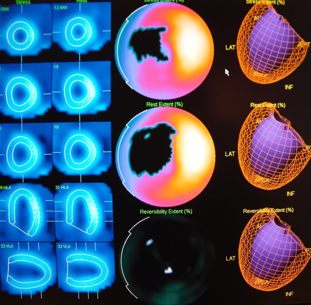
Radiopharmaceuticals can also be used to assess blood flow and tissue perfusion. Tc-99m-labelled compounds, such as Tc-99m Sestamibi, are employed in myocardial perfusion imaging to evaluate the blood supply to the heart muscle. Areas with reduced perfusion indicate regions of potential coronary artery disease.
Clinical Applications of Radiopharmaceutical Diagnostics
The use of radiopharmaceuticals in diagnostic imaging has revolutionised the field of medicine, providing valuable insights into a wide range of medical conditions. The following sections highlight some of the key clinical applications of radiopharmaceuticals.
Oncology
Radiopharmaceuticals play a critical role in detecting, staging, and monitoring cancer. FDG-PET imaging is widely used in oncology to detect primary tumours, assess the extent of metastasis, and monitor treatment response. New radiopharmaceuticals, such as Gallium-68 labelled peptides, are also being developed for specific tumour types, offering more targeted imaging options.
Cardiology
In cardiology, radiopharmaceuticals are employed to assess myocardial perfusion, detect coronary artery disease, and evaluate cardiac function. Myocardial perfusion imaging using Tc-99m-labelled compounds is a standard diagnostic tool for patients with suspected coronary artery disease, providing information on the severity and location of ischemia.
Neurology
Radiopharmaceuticals are also used to diagnose neurological conditions, including Alzheimer’s disease, Parkinson’s disease, and epilepsy. FDG-PET imaging can assess brain metabolism and detect areas of hypometabolism associated with neurodegenerative disorders. Additionally, radiolabelled dopamine transporter ligands are used to evaluate dopaminergic function in patients with Parkinsonian syndromes.
Infectious Diseases
Another significant application of radiopharmaceuticals is detecting infections. Tc-99m-labelled white blood cells and gallium-67 citrate are examples of radiopharmaceuticals used to localise sites of infection or inflammation in the body. These imaging techniques can be particularly useful in diagnosing osteomyelitis, prosthetic joint infections, and fever of unknown origin.
Safety and Regulation of Radiopharmaceuticals
Due to their radioactive nature, the safety and regulation of radiopharmaceuticals are of paramount importance. Stringent guidelines and protocols ensure the safe production, handling, and administration of these compounds.
Radiation Safety
The use of radiopharmaceuticals involves exposure to ionising radiation, which requires careful consideration of radiation safety principles. The effective dose of radiation received by the patient must be minimised while ensuring sufficient diagnostic accuracy. The selection of appropriate radiopharmaceuticals, optimised imaging protocols, and adherence to safety guidelines are crucial in reducing radiation exposure.
Regulatory Framework
Radiopharmaceuticals are subject to strict regulatory oversight to ensure their safety, efficacy, and quality. In the United Kingdom, the Medicines and Healthcare products Regulatory Agency (MHRA) regulates the production and use of radiopharmaceuticals. These regulations cover aspects such as the production facilities, quality control measures, and clinical use of radiopharmaceuticals.
Production and Distribution of Radiopharmaceuticals
The production and distribution of radiopharmaceuticals involve complex processes, including the production of radioactive isotopes, radiolabelling, and the formulation of the final product. These processes require specialised facilities and expertise.
Radioisotope Production
The production of radioactive isotopes, the core component of radiopharmaceuticals, typically occurs in nuclear reactors or cyclotrons. For example, Tc-99m is produced from the decay of Molybdenum-99 (Mo-99) in a generator system. The availability of these isotopes is critical to the continuous supply of radiopharmaceuticals for clinical use.
Radiolabelling and Formulation
Once the radioactive isotope is produced, it is combined with a carrier molecule to form the radiopharmaceutical. This process, known as radiolabelling, requires precision to ensure the stability and specificity of the compound. The formulated radiopharmaceutical must undergo rigorous quality control to verify its purity, radiochemical identity, and sterility before being distributed to healthcare facilities.
Emerging Trends and Future Directions
The field of radiopharmaceuticals is constantly evolving, with ongoing research and development aimed at improving diagnostic accuracy, expanding clinical applications, and enhancing patient safety. Several emerging trends and directions are shaping radiopharmaceuticals’ future in diagnostics.
Targeted Radiopharmaceuticals
There is a growing interest in developing radiopharmaceuticals that target specific molecular pathways or biomarkers associated with diseases. These targeted agents can provide more precise imaging and potentially enable early detection of diseases at a molecular level. Advances in molecular biology and imaging technologies are driving the development of these next-generation radiopharmaceuticals.
Theranostics
Theranostics, a combination of therapy and diagnostics, is an emerging field where radiopharmaceuticals are used for both diagnosis and treatment. For example, radiopharmaceuticals that target prostate-specific membrane antigen (PSMA) are being used for both imaging and targeted radionuclide therapy in prostate cancer. This approach allows for personalised treatment plans based on the imaging results.
Artificial Intelligence in Radiopharmaceutical Imaging
Artificial intelligence (AI) is increasingly being integrated into radiopharmaceutical imaging to enhance image analysis and interpretation. AI algorithms can assist in detecting subtle abnormalities, quantifying tracer uptake, and improving the accuracy of diagnostic reports. The integration of AI with radiopharmaceutical imaging has the potential to revolutionise clinical workflows and improve patient outcomes.
Conclusion
Radiopharmaceuticals have become indispensable tools in the field of nuclear medicine, providing unique insights into the functional and molecular aspects of diseases. Their application in diagnostic imaging has significantly advanced the ability to detect and monitor a wide range of medical conditions. As technology and research continue to evolve, radiopharmaceuticals are poised to play an even more significant role in personalised medicine, offering tailored diagnostic and therapeutic options for patients. The ongoing developments in targeted radiopharmaceuticals, theranostics, and AI integration hold promise for further improving the accuracy and efficacy of diagnostic imaging, ultimately enhancing patient care.
In conclusion, the future of radiopharmaceuticals in diagnostics is bright, with ongoing advancements promising to expand their clinical utility and improve patient outcomes. As the field continues to innovate, radiopharmaceuticals will remain at the forefront of diagnostic imaging, helping to unlock new possibilities in the early detection and treatment of diseases.
Disclaimer
This article is intended for informational and educational purposes only. It does not constitute medical, scientific, or professional advice and should not be relied upon as such. While every effort has been made to ensure the accuracy and currency of the content at the time of publication, Open Medscience makes no representations or warranties, express or implied, regarding the completeness, reliability, or suitability of the information contained herein.
Readers are advised to consult qualified healthcare professionals or specialists before making any decisions based on the information provided. The use of radiopharmaceuticals is subject to strict medical supervision, regulatory oversight, and professional judgement, and may not be appropriate or available in all clinical settings.
Open Medscience disclaims any liability for any loss, damage, or injury arising from the use of or reliance on this article or any of its contents. The views expressed are those of the authors and do not necessarily reflect the official policy or position of Open Medscience or its affiliates.
home » diagnostic medical imaging blog » nuclear medicine imaging »


