Summary: This article examines imaging techniques that utilise radioactive substances to visualise internal biological processes. It outlines the principles underlying radiographic methods such as Positron Emission Tomography (PET), Single Photon Emission Computed Tomography (SPECT), and autoradiography. Additionally, the article explores contemporary advancements in radioimaging technology, highlighting significant applications in medicine, research, and industrial fields, while also considering the safety of radiation and its ethical implications.
Keywords: Radioactivity, PET imaging, SPECT imaging, Autoradiography, Medical imaging, Radiation safety
Fundamentals of Radioactive Imaging
Radioactivity has been a cornerstone of imaging techniques in medical diagnostics and scientific research for over a century. Imaging techniques that employ radioisotopes are invaluable for their ability to reveal functional and biochemical activity within living organisms. By using radioactive isotopes as tracers, clinicians and scientists can observe metabolic processes, diagnose disease, and monitor treatment effectiveness.
This article focuses primarily on three distinct imaging techniques that utilise radioactivity: Positron Emission Tomography (PET), Single Photon Emission Computed Tomography (SPECT), and autoradiography, examining each method’s theoretical basis, practical applications, and recent technological advancements.
Radioactive imaging relies upon isotopes, unstable atoms that emit radiation as they decay to more stable forms. These emissions—alpha, beta, gamma, or positrons—are detected by specialised imaging equipment to produce diagnostic images or scientific data.
Radiotracers and Their Properties
Radiotracers, isotopically labelled compounds introduced into the body or materials, are fundamental in radioactive imaging. For medical purposes, these compounds typically have short half-lives to minimise radiation exposure, yet are sufficiently long to capture the required imaging data. Examples include fluorine-18, used in PET imaging, and technetium-99m, which is extensively used in SPECT imaging.
Positron Emission Tomography (PET)
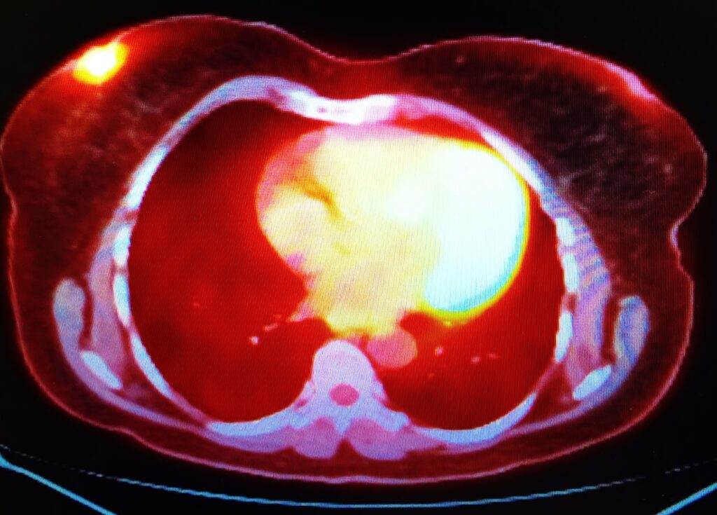
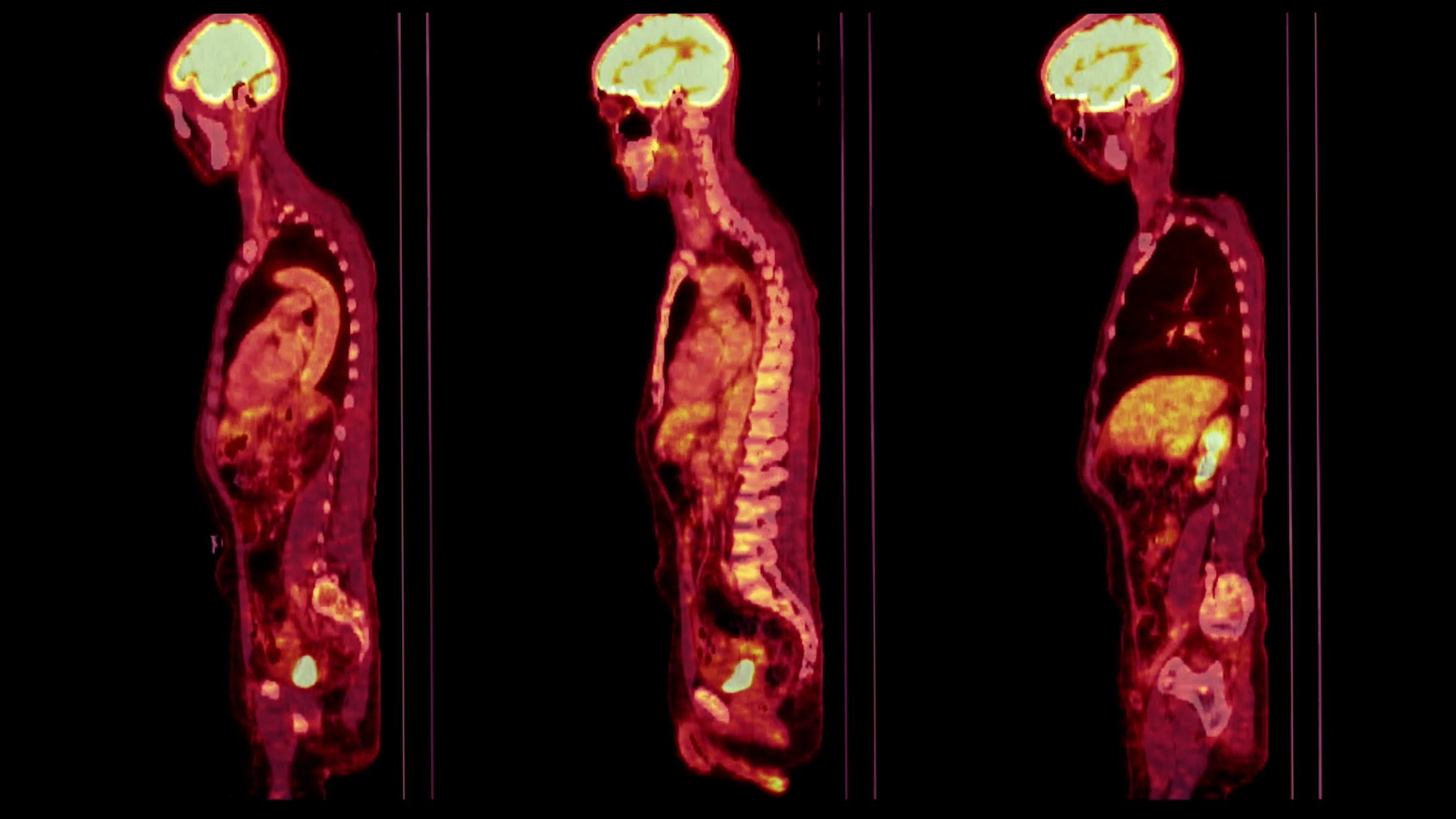
PET imaging utilises radioisotopes that emit positrons, positively charged electrons that annihilate with electrons upon encountering them, producing two gamma photons that move in opposite directions. Detection of these photons simultaneously enables the precise mapping of radioactive decay locations, providing detailed functional imagery.
Principles and Instrumentation
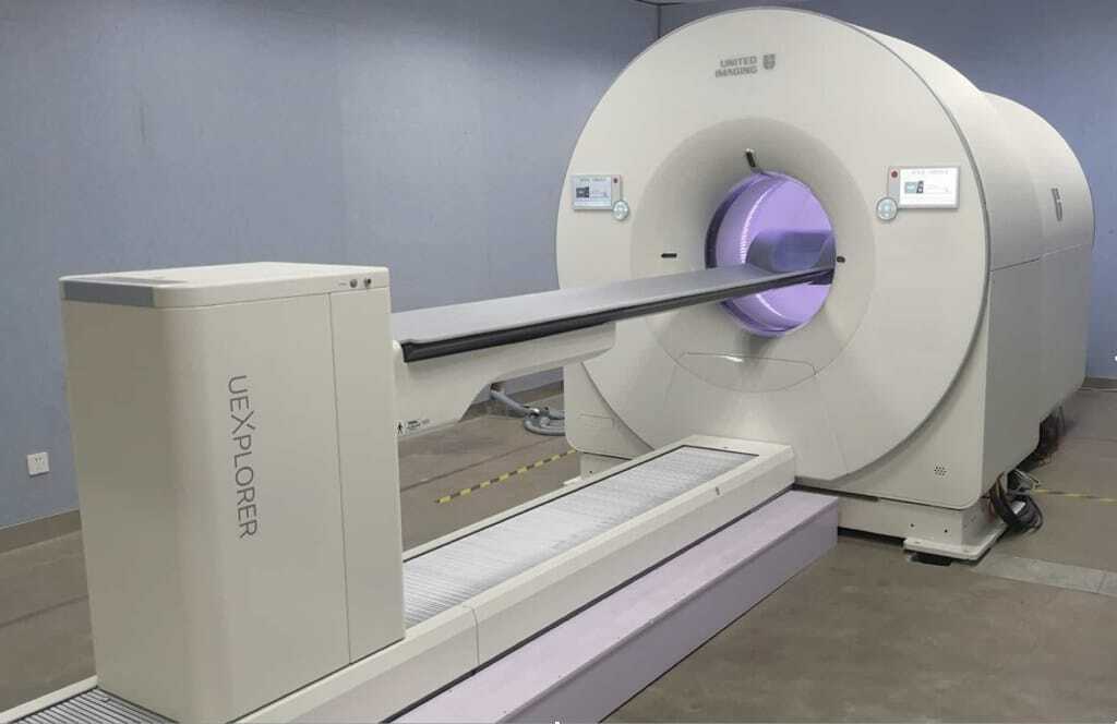
PET scanners consist of ring-shaped detectors capturing gamma photons emitted during positron annihilation. Algorithms reconstruct the gamma-ray signals into detailed, three-dimensional images, allowing clinicians to visualise metabolic processes in organs such as the brain, heart, and tumours.
Clinical Applications
PET imaging has a significant impact on oncology, neurology, and cardiology. In oncology, radiolabelled glucose (18F-FDG) highlights metabolically active cancer cells. Neurologically, PET detects conditions such as Alzheimer’s and Parkinson’s diseases through characteristic metabolic patterns. Cardiology benefits from PET by assessing myocardial blood flow and metabolism.
Recent Advances
Recent innovations include hybrid PET-MRI and PET-CT scanners that integrate anatomical with functional imaging. Hybrid systems enhance diagnostic accuracy, reduce imaging times, and provide comprehensive datasets crucial for personalised medicine approaches.
Single Photon Emission Computed Tomography (SPECT)
Unlike PET, SPECT employs isotopes that emit single gamma photons directly, without the need for positron intermediates. Commonly used isotopes in SPECT imaging include technetium-99m, iodine-123, and thallium-201.
Mechanism and Technology
Gamma cameras rotate around the patient, capturing photon emissions from various angles. Images are reconstructed into a three-dimensional representation of the radiotracer distribution. Compared to PET, SPECT generally offers lower resolution but is more widely available and less costly.
Clinical and Research Uses
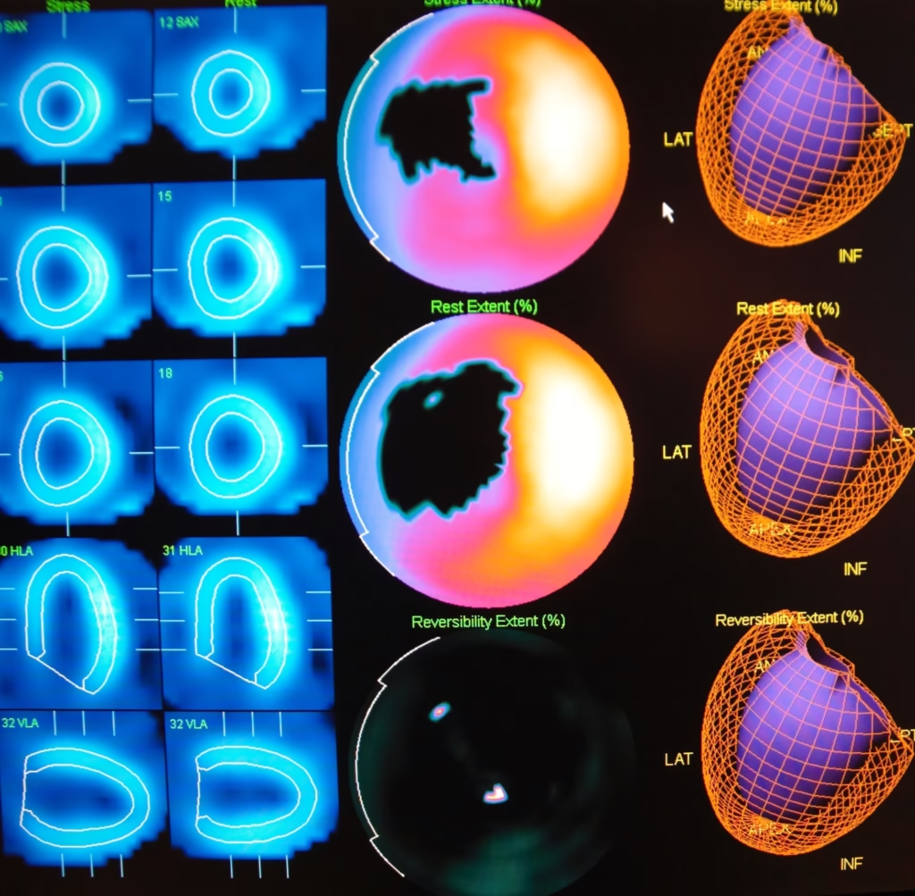
SPECT imaging is widely used in cardiology for myocardial perfusion imaging, which identifies areas of reduced blood supply indicative of coronary artery disease. It is equally valuable in neurology for diagnosing epilepsy and stroke, as well as in endocrinology, particularly for thyroid imaging using radioactive iodine isotopes.
Innovations and Developments
Recent developments in SPECT include advancements in detector technology, improving spatial resolution and sensitivity. New collimator designs and novel radiotracers offer enhanced diagnostic accuracy and broader clinical applications.
Autoradiography
Autoradiography is a method used primarily in research rather than clinical practice, visualising radioactive substances distributed in biological tissues or cellular samples.
Technique and Application
In autoradiography, thin tissue sections or gels are placed against photographic or phosphor screens, which record radioactive emissions to create a visual map of tracer distribution. This technique is essential in pharmacology, molecular biology, and neuroscience research, providing detailed insights into molecular pathways, receptor binding, and enzyme activities.
Current Trends
Advances in digital autoradiography systems offer higher-resolution images and quantitative analyses, significantly benefiting biomedical research, particularly in drug discovery and cellular biology.
Radiation Safety and Ethical Considerations
Using radioactive substances necessitates rigorous safety and ethical standards. Regulatory bodies enforce strict guidelines to minimise radiation exposure to patients, researchers, and the environment.
Dose Management and Safety Protocols
Modern imaging methods aim for the lowest radiation dose possible while maintaining diagnostic efficacy. Advanced software algorithms reduce scanning durations, thus decreasing radiation exposure. Radiotracers with shorter half-lives and improved targeting efficiency also help mitigate risks.
Ethical Challenges
Ethically, radioactivity-based imaging raises concerns regarding informed consent, particularly regarding cumulative radiation exposure from multiple imaging sessions. Ethical committees continually review and update guidelines to strike a balance between medical benefits and potential risks.
Industrial and Environmental Applications
Beyond medicine and biological research, radioactive imaging extends to industrial and environmental fields. Techniques such as gamma radiography offer non-destructive testing (NDT) methods for assessing structural integrity in engineering, aerospace, and materials science. Environmental monitoring using radioactive tracers helps detect the spread of contamination and track the movement of water resources.
Future Directions
Radioactive imaging technology continues to evolve, driven by the demand for precision diagnostics and treatment monitoring. Future developments will focus on greater sensitivity, faster imaging, and personalised radiotracers, enhancing imaging efficacy across clinical and research settings.
Innovations integrating artificial intelligence (AI) and machine learning promise improved image interpretation and predictive diagnostics. Emerging radiopharmaceuticals and advanced detector technologies will enhance diagnostic precision and reduce patient exposure.
Conclusion
Imaging techniques that employ radioactivity remain fundamental in modern medicine and research, providing unparalleled insights into physiological processes. PET, SPECT, and autoradiography each offer unique capabilities, underpinning critical advancements in diagnosis, treatment monitoring, and scientific discovery. Continuous technological and methodological improvements promise increasingly precise, safe, and ethical imaging solutions, ensuring radioactive imaging remains central to medical, industrial, and environmental innovation.
Disclaimer
The content provided in this article, Unlocking Life’s Secrets: The Power and Potential of Radioactive Imaging, is intended for informational and educational purposes only. While every effort has been made to ensure accuracy and scientific rigour, the information should not be construed as medical advice, diagnostic guidance, or a substitute for professional consultation with qualified healthcare or scientific professionals.
Any references to specific techniques, technologies, or radiopharmaceuticals are for illustrative purposes and do not imply endorsement by Open Medscience. The safety, ethical considerations, and regulatory requirements surrounding the use of radioactive substances must be adhered to in accordance with local and international guidelines.
Open Medscience disclaims any liability arising directly or indirectly from the use or interpretation of the material presented. Readers are encouraged to seek expert advice when applying the concepts discussed in clinical, research, or industrial contexts.
home » blog » nuclear medicine imaging »