
From Pixels to Patterns: Big Data and AI Transforming Medical Imaging
 By
By
Big Data will be the foundation for personalised healthcare, especially the application of algorithmic tools capable of converting raw data to large datasets.
By
Big Data will be the foundation for personalised healthcare, especially the application of algorithmic tools capable of converting raw data to large datasets.
 By
By
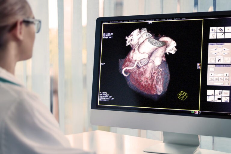
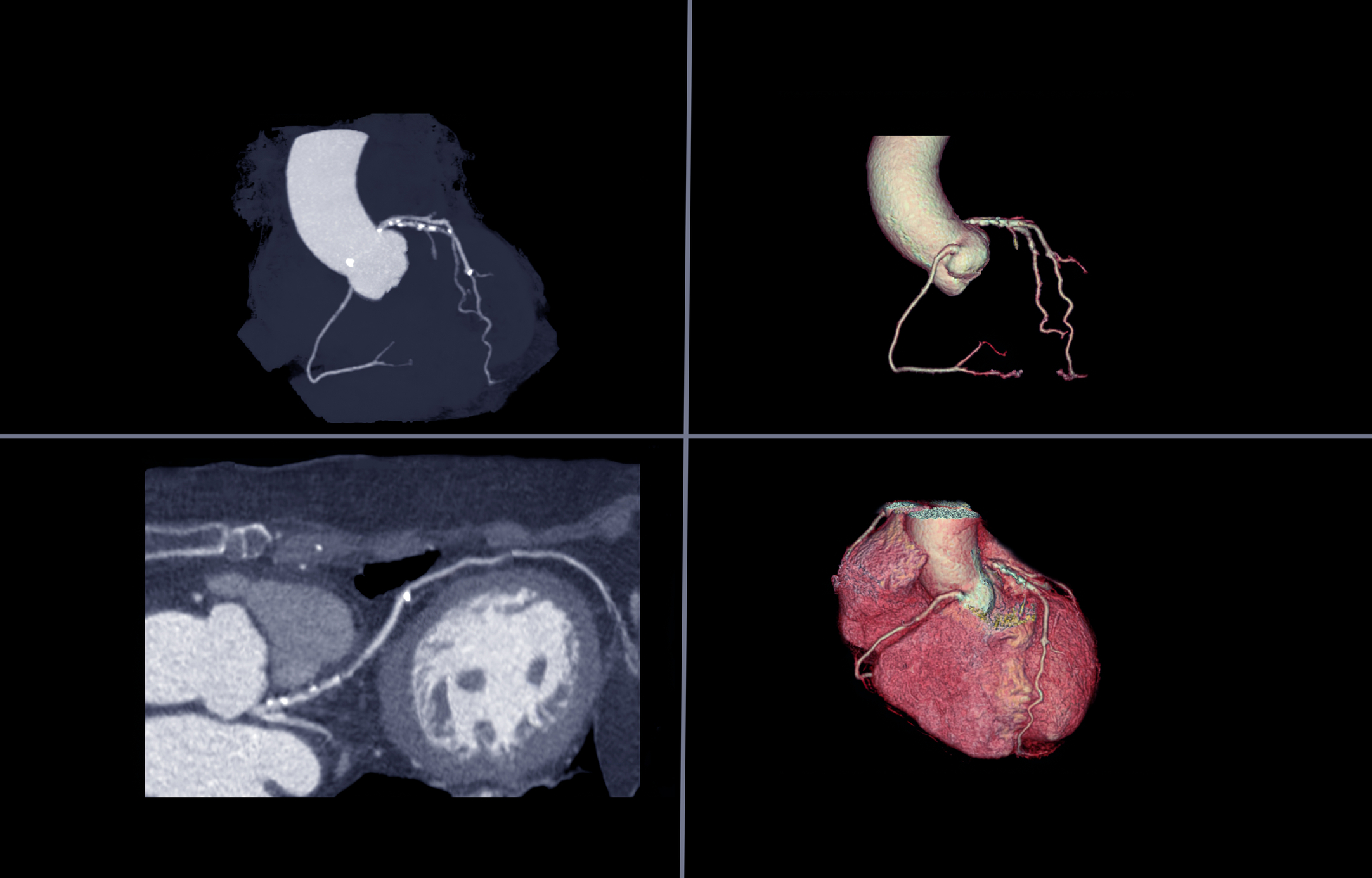
The new technologies emerging in the clinical setting include fractional flow reserve (FFR)-CT, CT perfusion imaging and coronary plaque assessment. Image for illustration only. Person depicted is a model.
 By
By
CTCA imaging has revolutionised how physicians detect coronary artery disease due to its exceptional sensitivity.
 By
By
Modern medical lasers are used in various clinical applications, including cancer therapy and ophthalmology.
 By
By
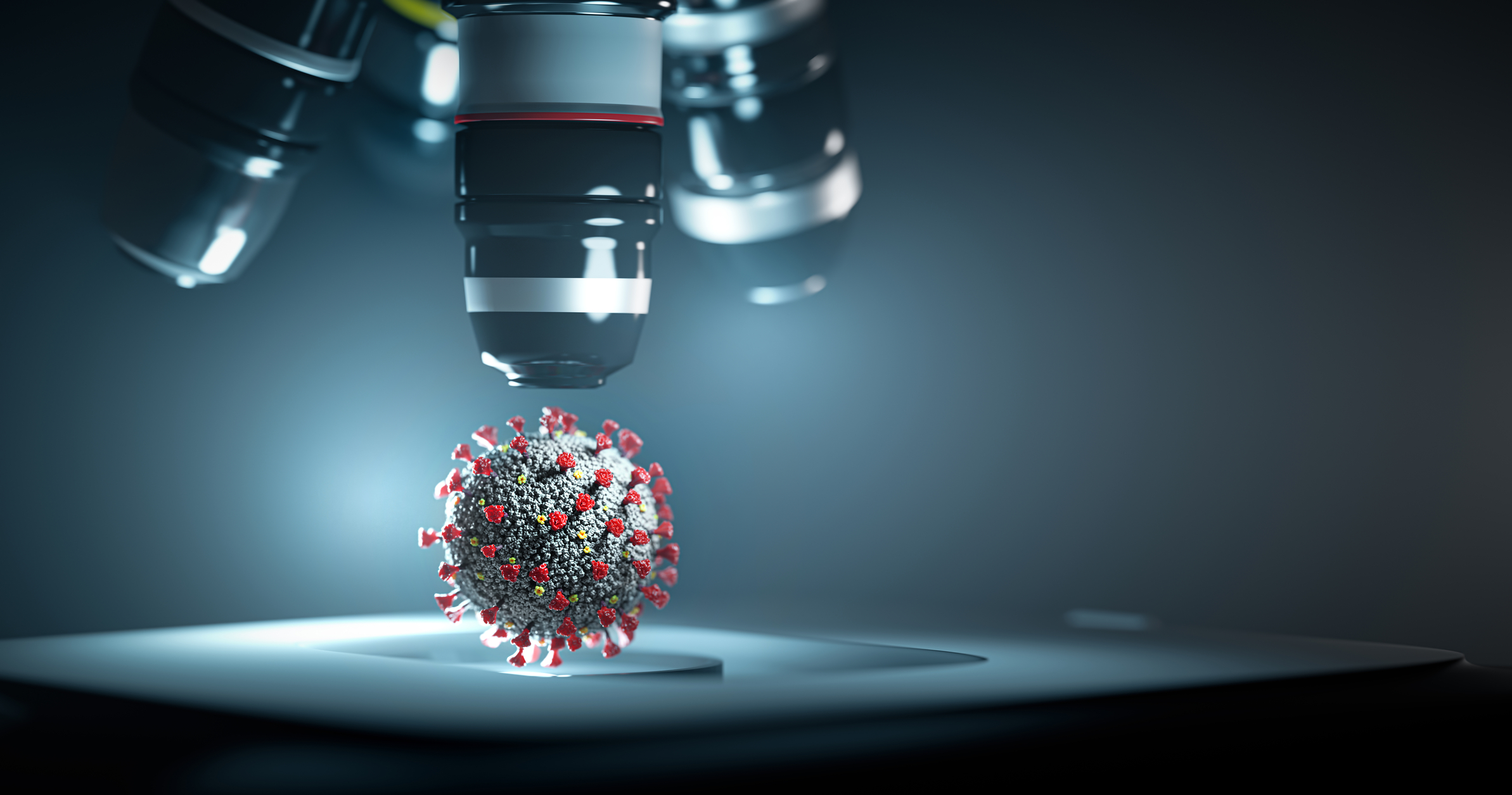
The structures of a virus can be elucidated by using the high resolving power of scanning electron microscopy.
 By
By
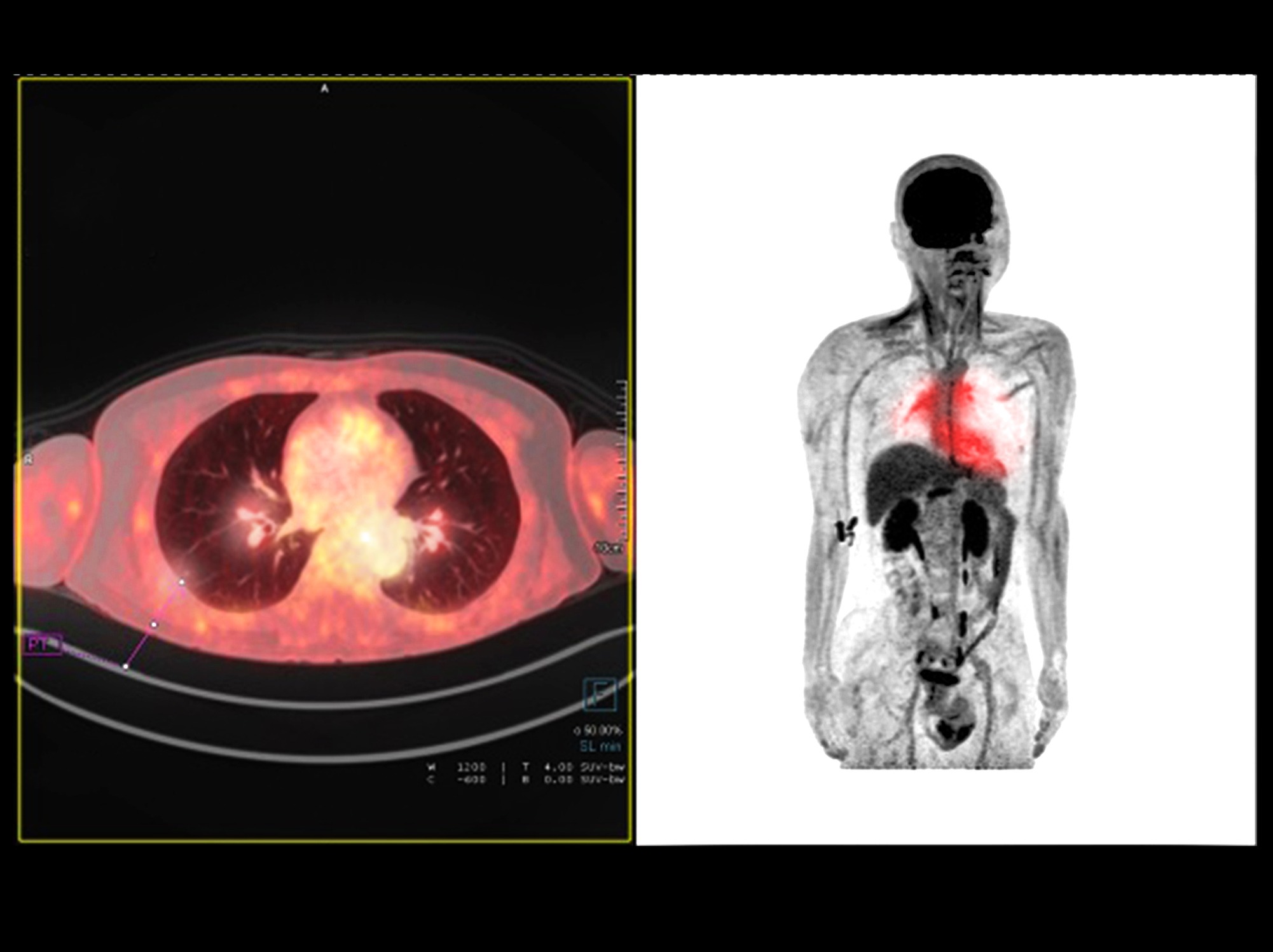
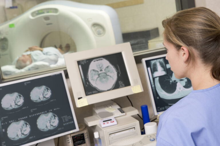
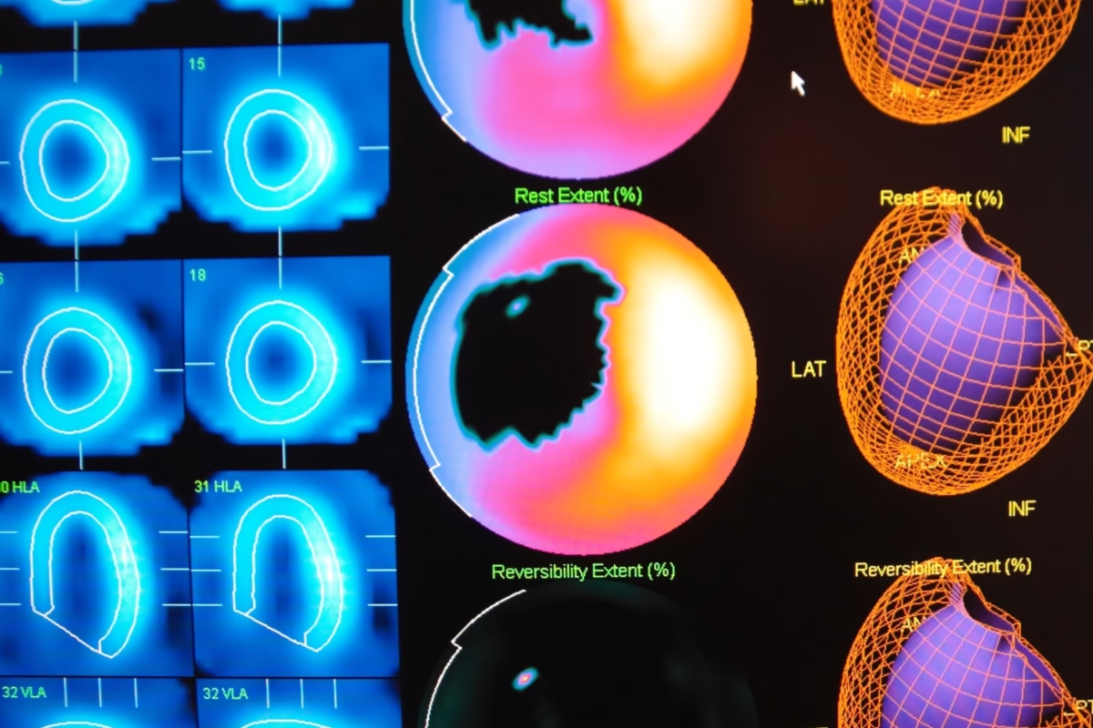
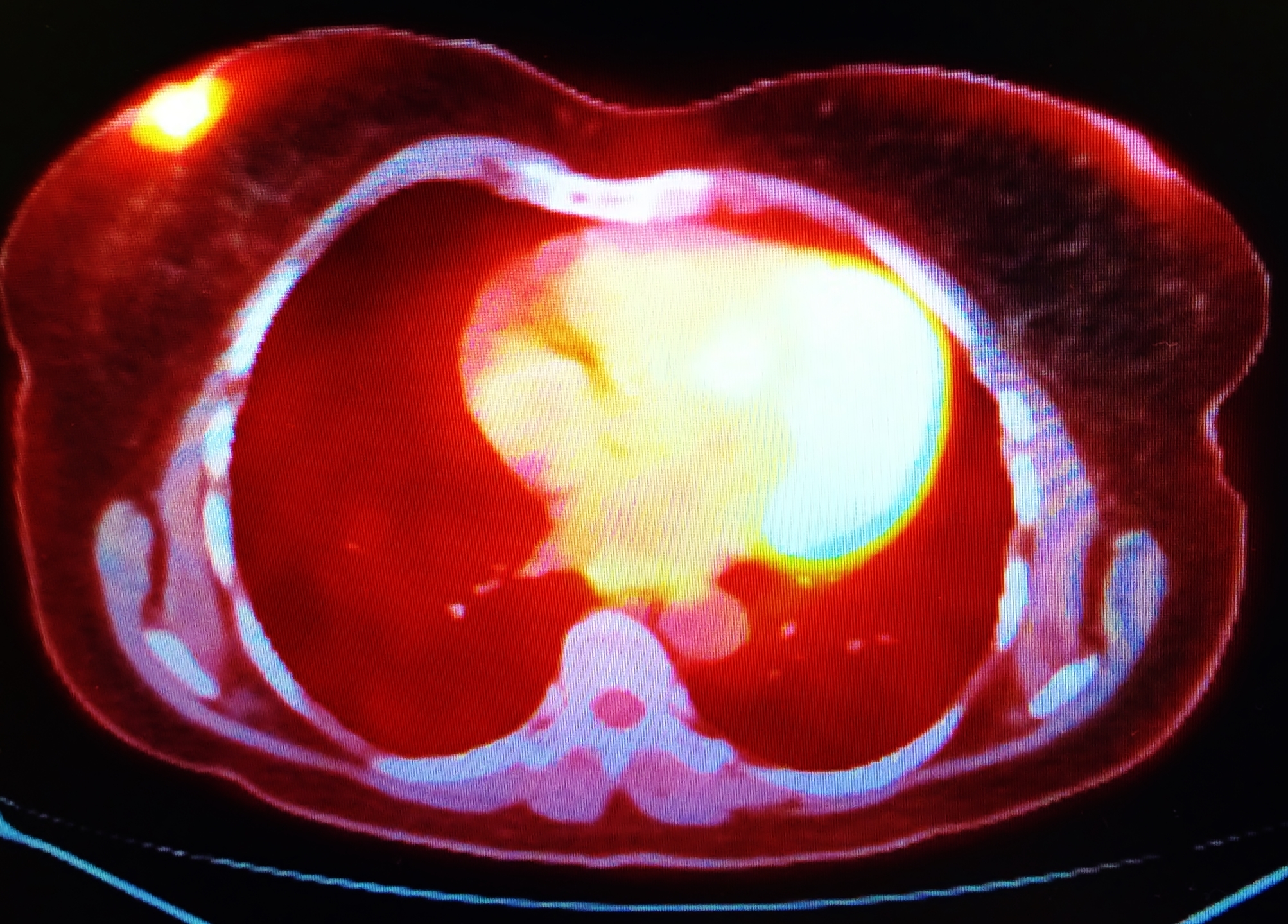
PET imaging is used in oncology, neurology and cardiology.
 By
By
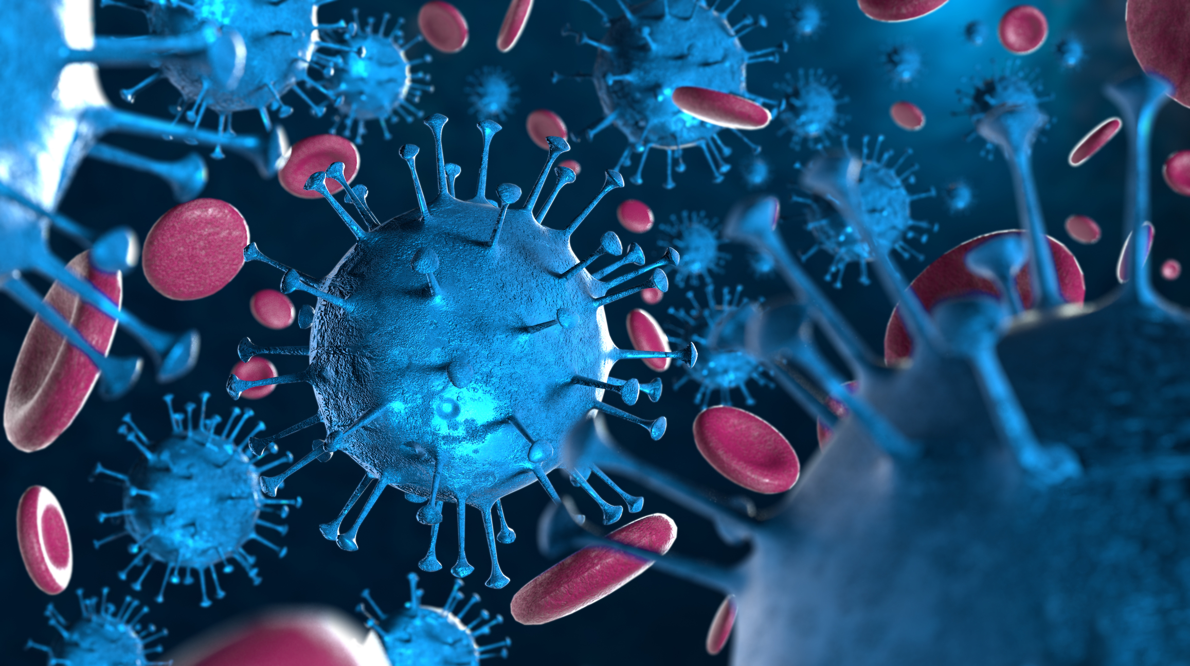
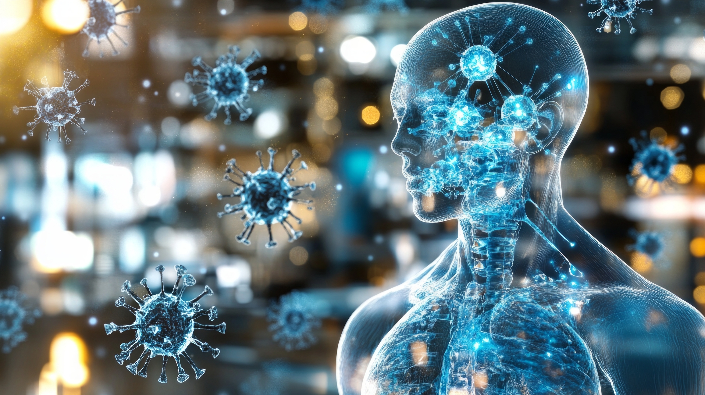
COVID-19 is caused by coronavirus virions that are enveloped spherical shaped virus crown spikes.
 By
By
WannaCry infects computers and encrypts window files on the hard drive, making them impossible for users to access.
 By
By
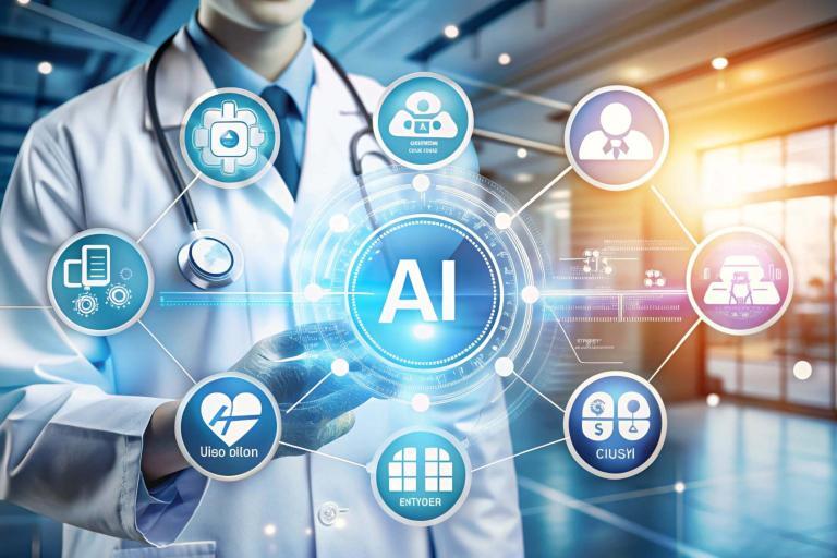
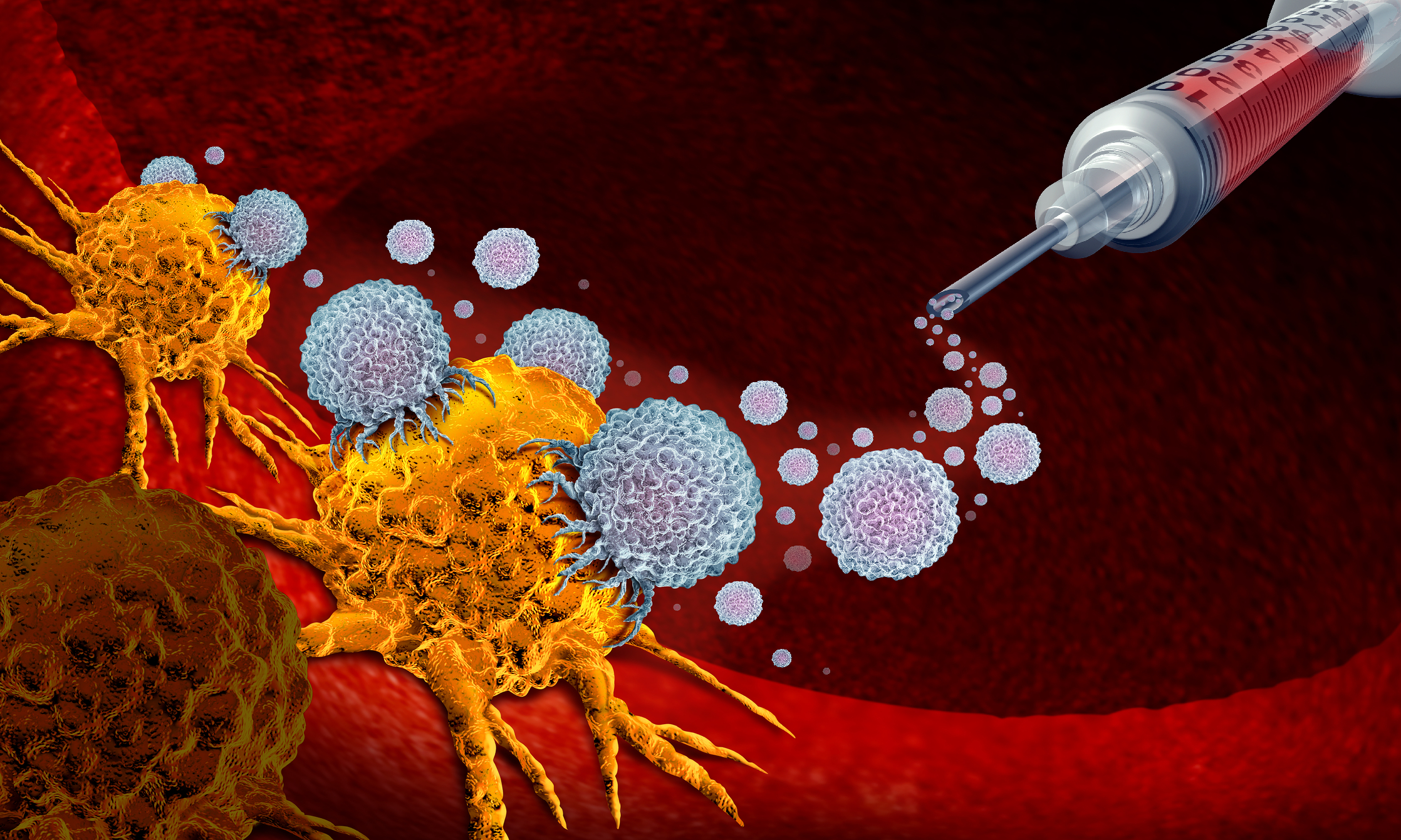
Artificial intelligence (AI) and the study of algorithms, known as machine learning, will analyse complex medical imaging data from patients.
 By
By
Medical imaging plays a vital role in the early detection of breast cancer including those with BRCA1 or BRCA2 mutations. Image for illustration only. Person depicted is a model.
 By
By
Imaging agents can be used to evaluate organ function, detect cancer, measure blood flow and follow metabolic processes.
 By
By
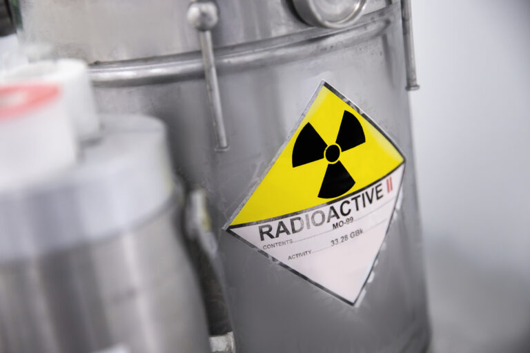
Radiopharmaceuticals are used in nuclear medicine for the application of medical imaging and therapy.
 By
By
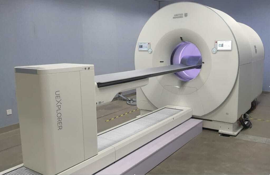
EXPLORER, the world’s first medical imaging scanner to produce a 3-D picture of the whole human body.
 By
By
Conventional X-ray systems are based on an immovable X-ray tube whereas the CT scanner uses a rotational X-ray source. Image for illustration only. People depicted are models.
 By
By
These cancer destroying machines are capable of providing proton beam therapy via pencil beam scanning.
 By
By
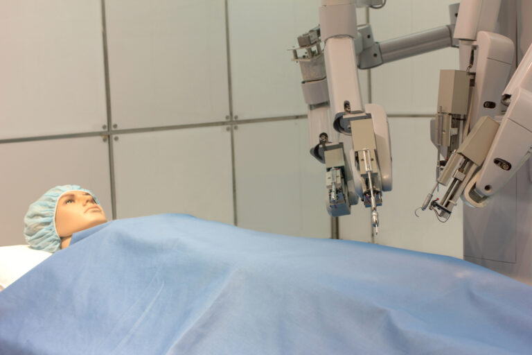
The da Vinci Surgery System is the most universal robot used in robotic surgery systems.
 By
By
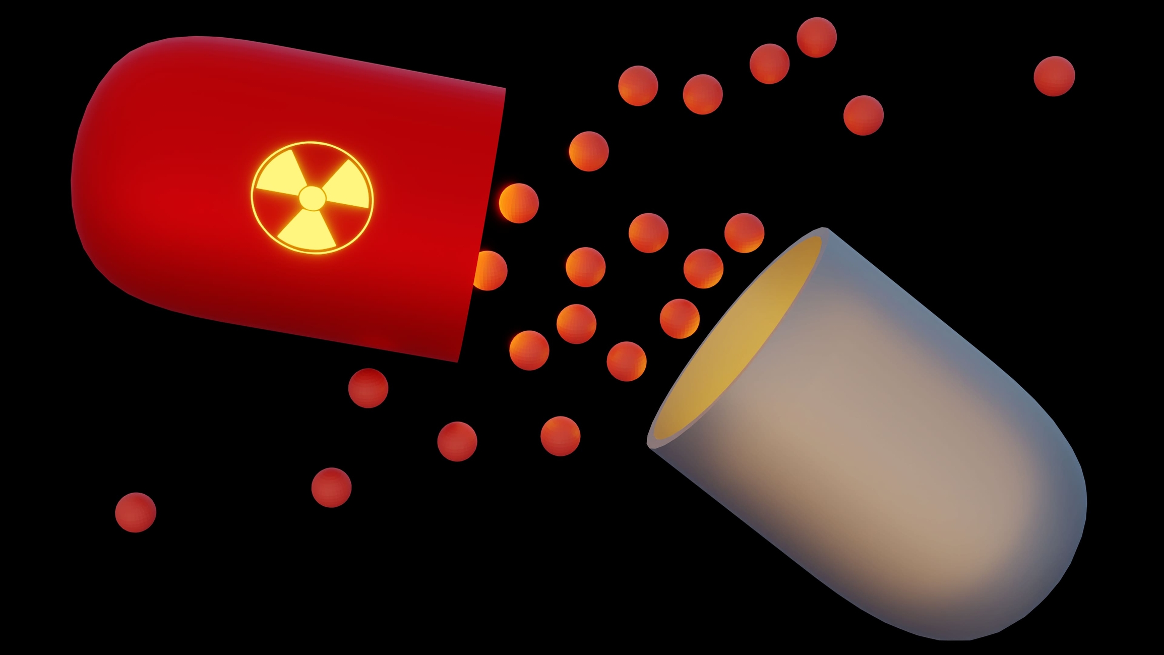
Targeted radionuclide therapy was first used to treat cancer for an ‘over-active’ thyroid using radioactive iodine-131 seeds.
 By
By
The most commonly used medical radioisotope in diagnostic procedures is technetium-99m.
 By
By
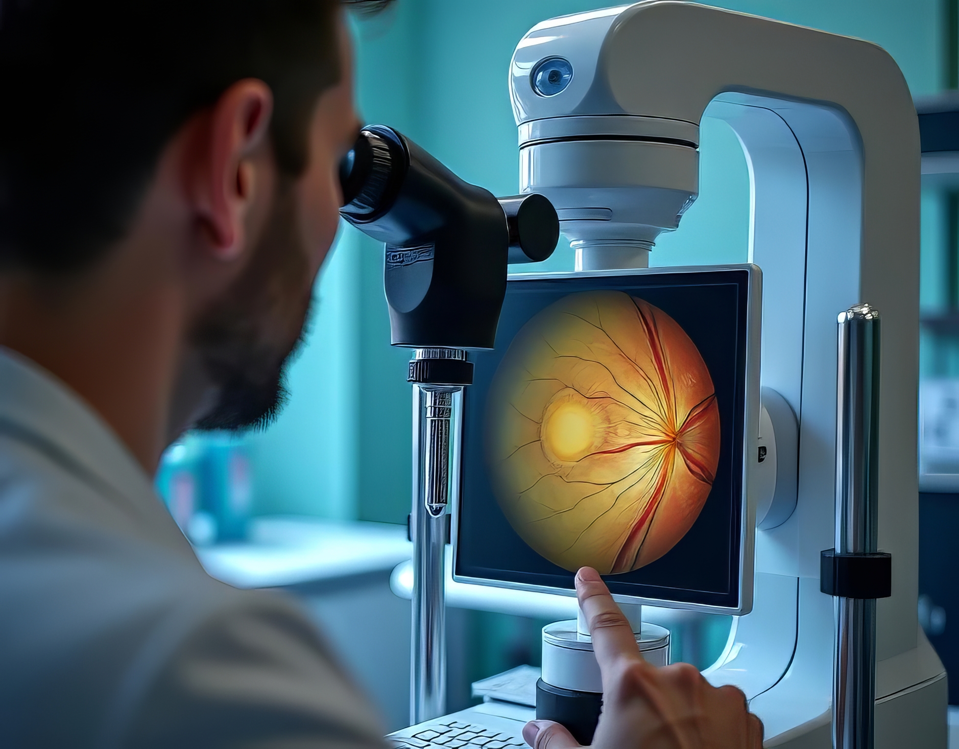
Since the 1800s, optic disc photography has been considered the gold standard for optic nerve evaluation. Image for illustration only. Person depicted is a model.
 By
By
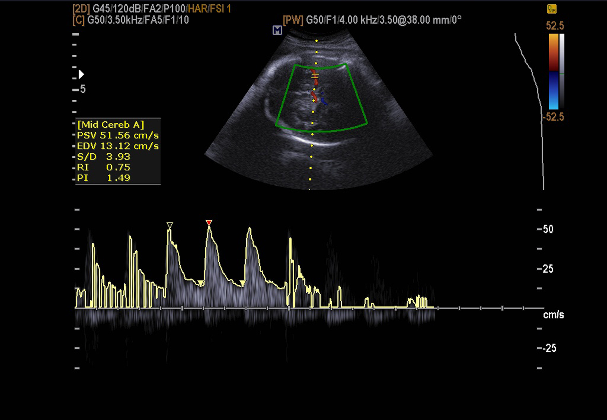
The magic of ultrasound imaging enables healthcare professionals to look inside the human body without being invasive.
 By
By
Artificial Intelligence will play a vital role in the analysis of vasts amounts of medical imaging data.
 By
By
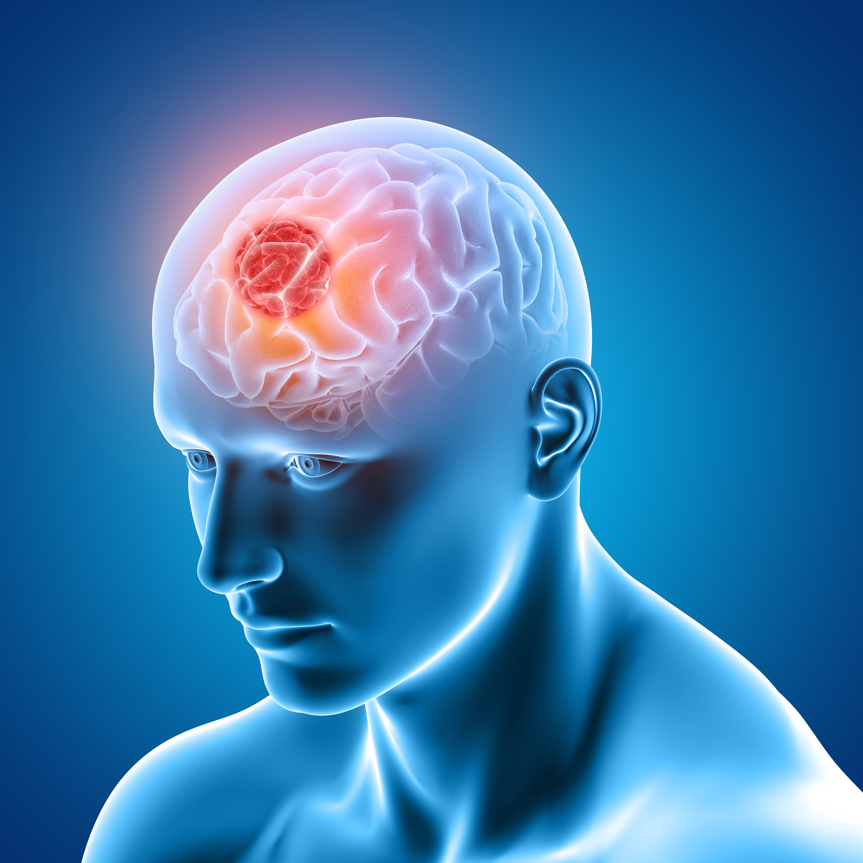
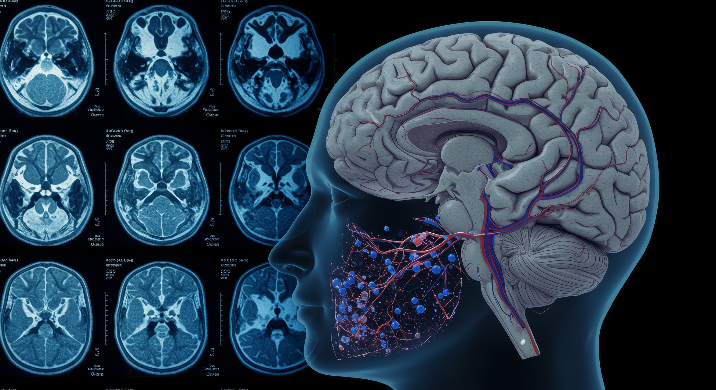
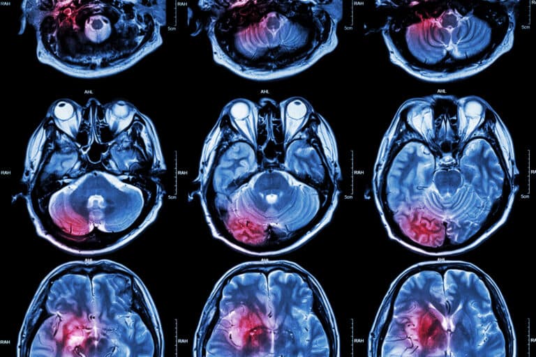
A useful brain imaging technique uses functional magnetic resonance imaging to analyse metabolic changes such as blood oxygenation.
 By
By
These non-radioactive labels can be incorporated into small molecules to study in vivo metabolic pathways in real-time.
 By
By
The diagnostic breast imaging tool Positron Emission Mammography uses short-lived positron isotopes to detect breast cancer.
 By
By
Brachytherapy techniques have been a powerhouse in the treatment of cancer since the beginning of the twentieth century.