Bridging the Gap Between Medical Imaging and Neurological Care
 By
By
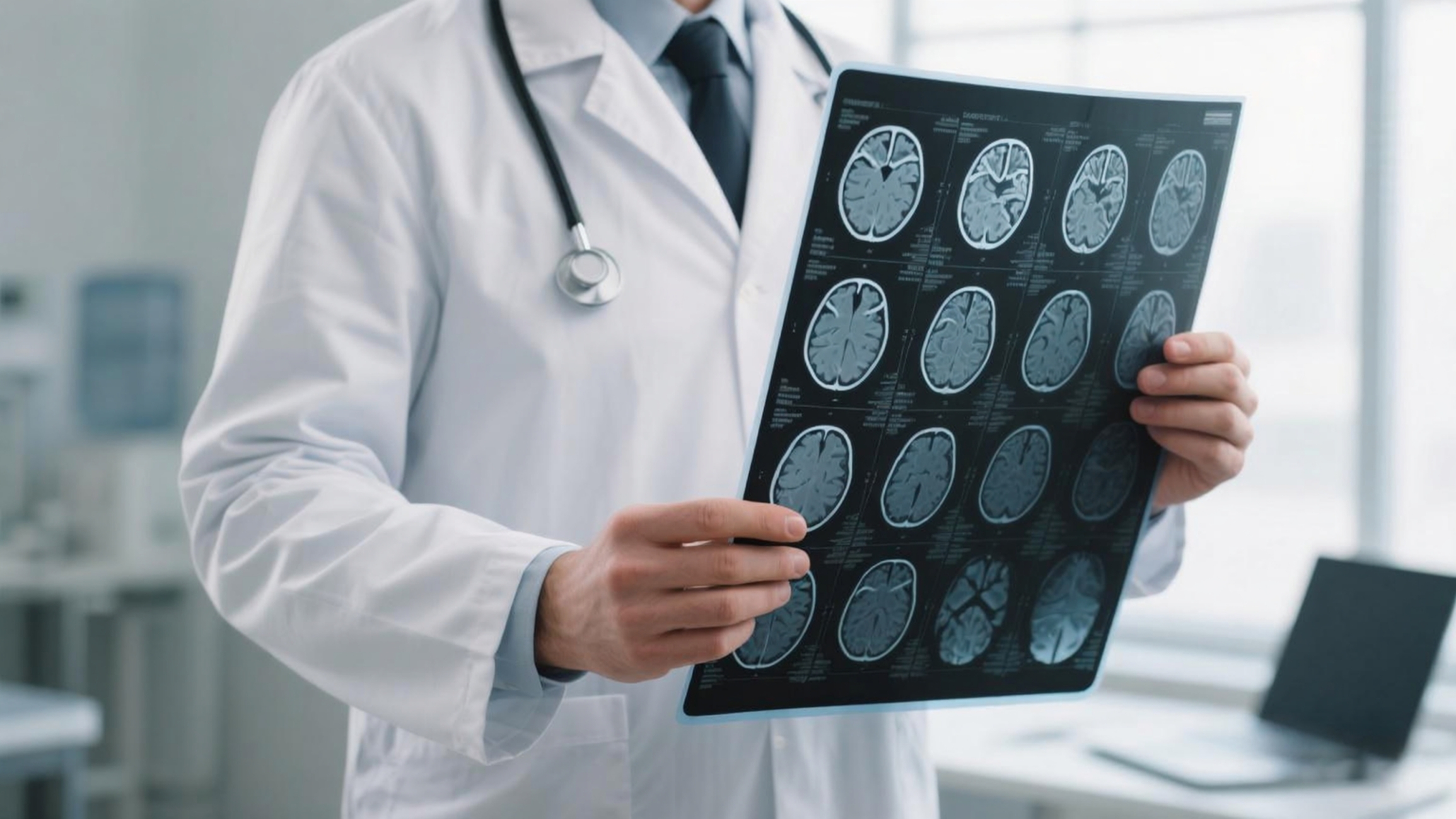
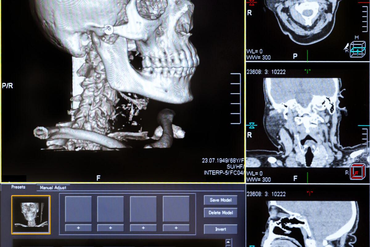
Discover the complexities of neurological care and the necessity for seamless information transfer in clinical practice.
By
Discover the complexities of neurological care and the necessity for seamless information transfer in clinical practice.
 By
By
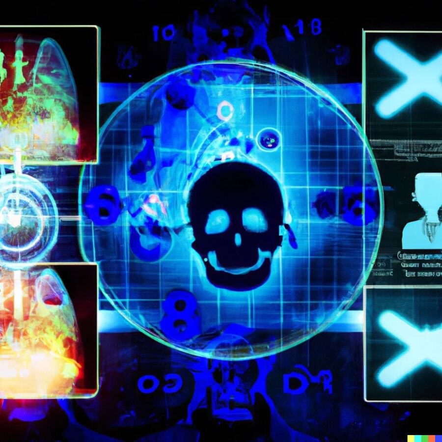
Learn about Modern Medical Imaging and Radiation Therapy and how advanced technology shapes the future of oncological treatment.
 By
By
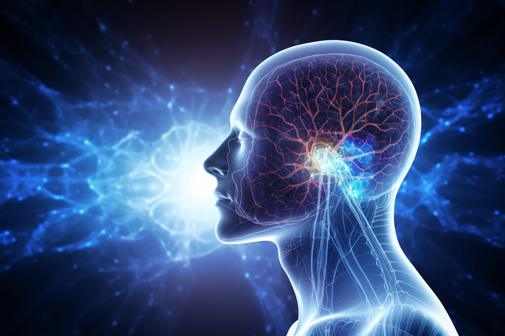
Stay updated on medical imaging breakthroughs that are transforming healthcare and improving patient outcomes in innovative ways.
 By
By
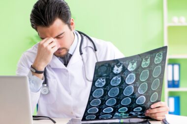
Learn about radiology negligence and the importance of accuracy in imaging to prevent delays in diagnosis and treatment. Image for illustration only. Person depicted is a model.
 By
By
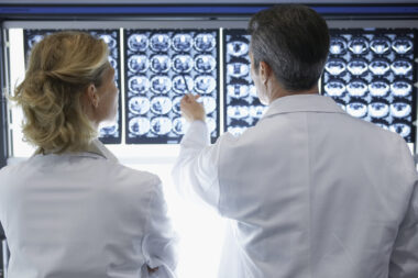
Discover the role of radiology and medical imaging in modern healthcare and how they enhance clinical decision-making. Image for illustration only. People depicted are models.
 By
By
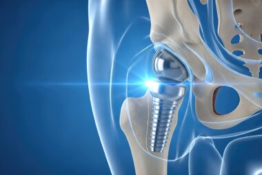
Discover how hip replacement imaging has advanced, improving diagnosis and surgical planning for better mobility outcomes.
 By
By
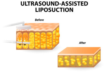
Find out how liposuction options can support your journey towards higher self-esteem and holistic body confidence enhancement.
 By
By
Discover the importance of musculoskeletal imaging diagnosis for accurate treatment of joint and back pain condition. Image for illustration only. People depicted are models.
 By
By
Explore ways to ensure your lower body healthy. Our article provides strategies to enhance flexibility and strength effectively. Image for illustration only. People depicted are models.
 By
By
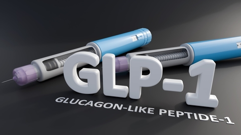
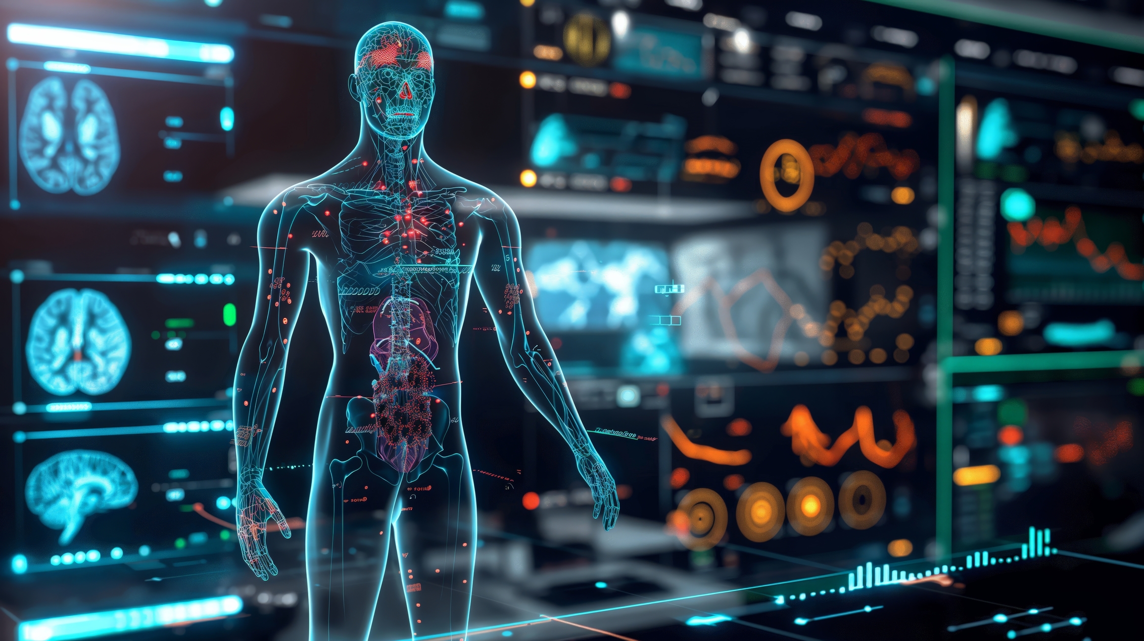
Discover the transformative GLP-1 imaging effects on fat distribution and inflammatory patterns in modern healthcare.
 By
By
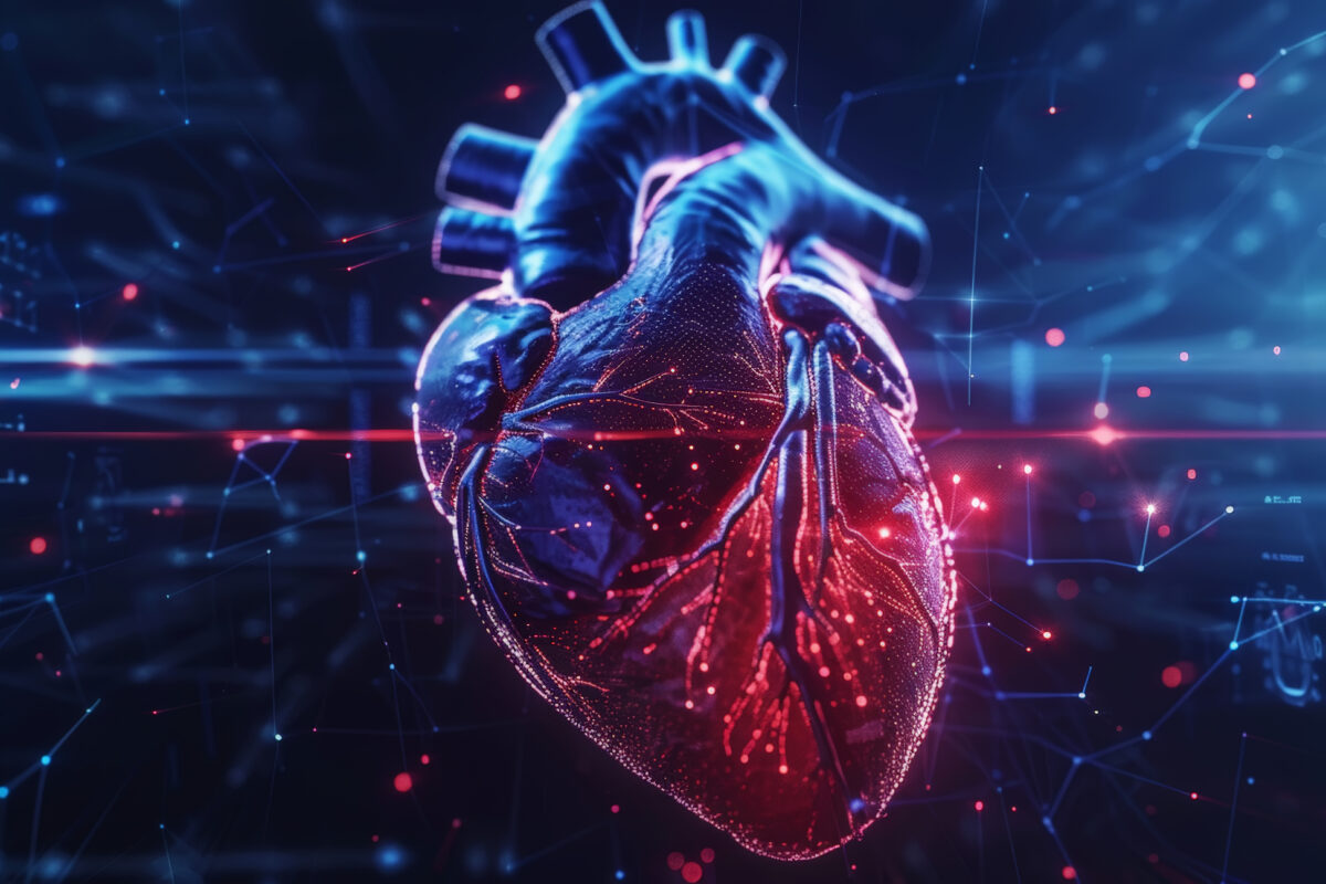
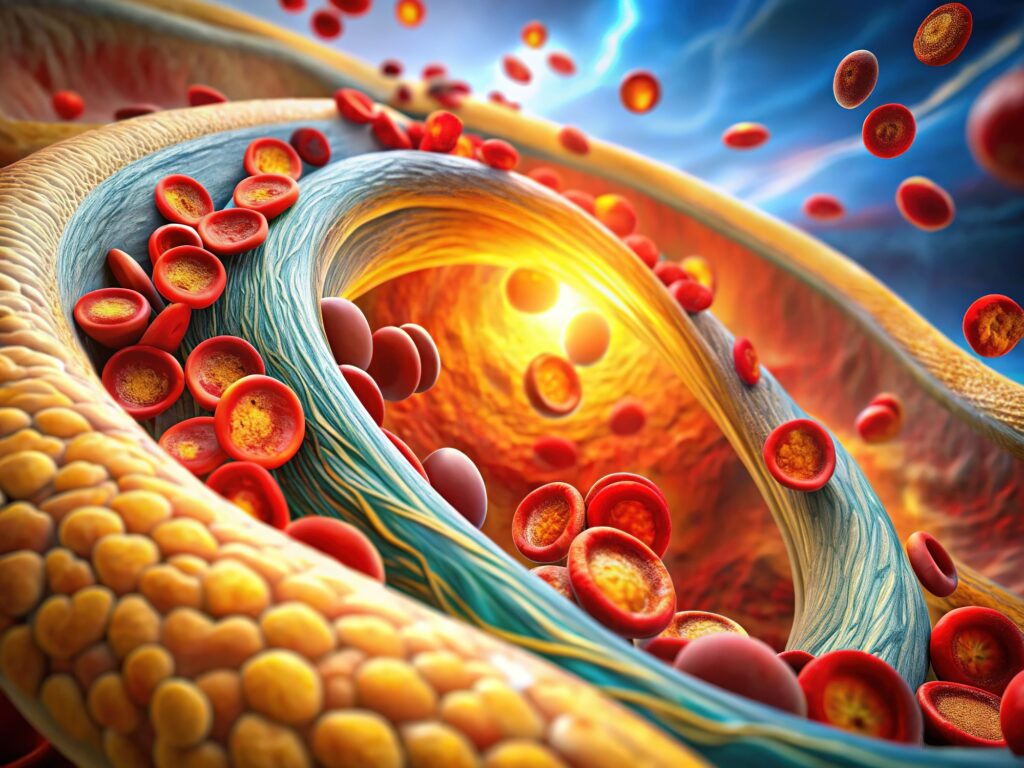
Discover how heart imaging is transformed by new technology, enhancing the diagnosis and treatment of cardiovascular disease.
 By
By
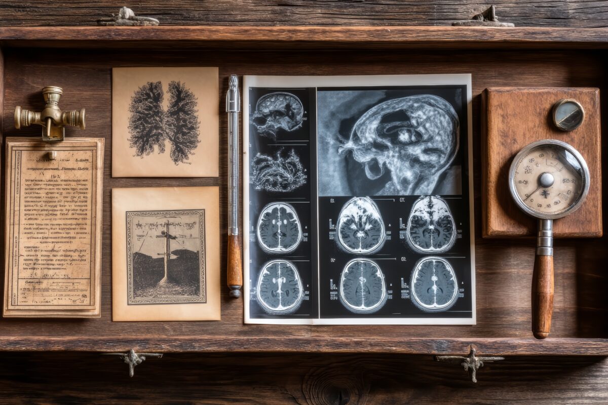
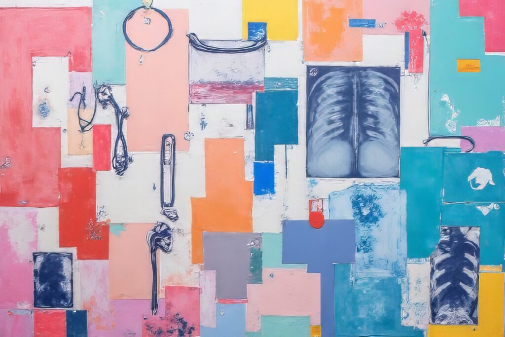
Explore the groundbreaking work of the pioneers of medical imaging and how they changed the landscape of modern medicine.
 By
By
Discover how quantum wellness technology enhances human vitality by utilizing principles from quantum physics and bioenergetics.
 By
By
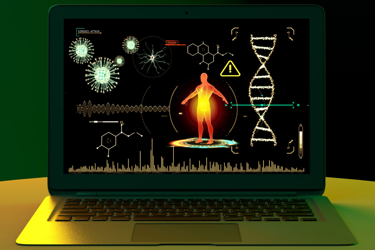
Discover how medical imaging for viral detection is revolutionizing diagnostics, offering insights into viral infections and treatments.
 By
By
Learn key new nurse lessons to better prepare for your first job and anticipate the challenges ahead in nursing.
 By
By
Discover the fascinating types of medical imaging, from X-rays to advanced molecular techniques, shaping health care today.
 By
By
Discover how Zepbound weight management can help achieve significant weight loss and improve metabolic conditions like diabetes.
 By
By
Understand the role of MRI carotid inflammation as a PET-equivalent method for detecting plaque inflammation in strokes.
 By
By
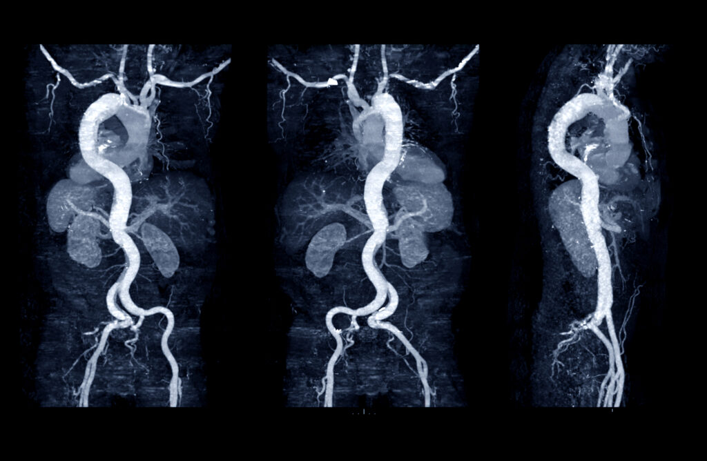
Discover the significance of the Aneurysm Progression Marker in predicting AAA growth with non-invasive perivascular fat density measurements.
 By
By
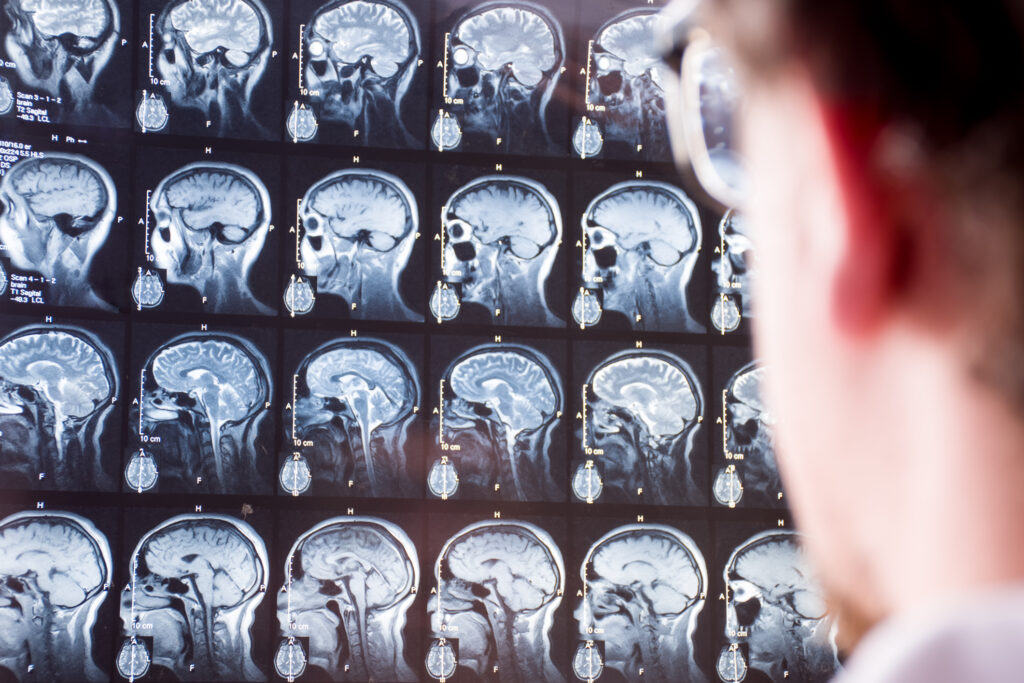
Learn how functional MRI technology is used to assess brain injury. Understand the complexities of mTBI symptoms and recovery.
 By
By
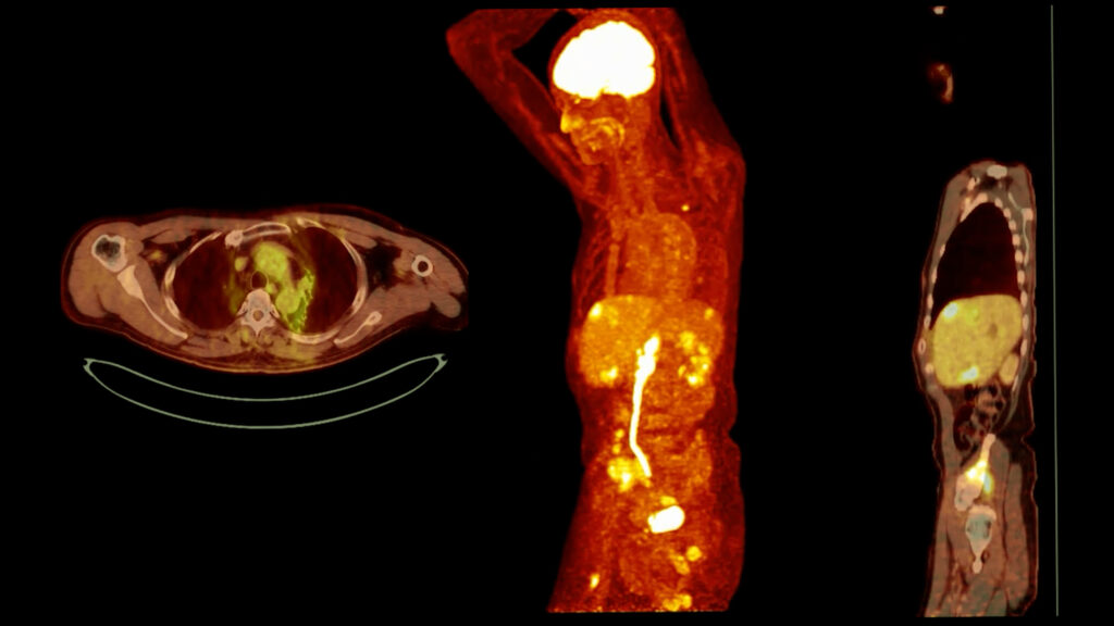
Understand the importance of PET imaging in healthcare, from cardiac assessments to patient journeys through nuclear medicine.
 By
By
Evaluate your expertise with the medical imaging quiz, focusing on imaging parameters, radiation safety, and practical case studies.
 By
By
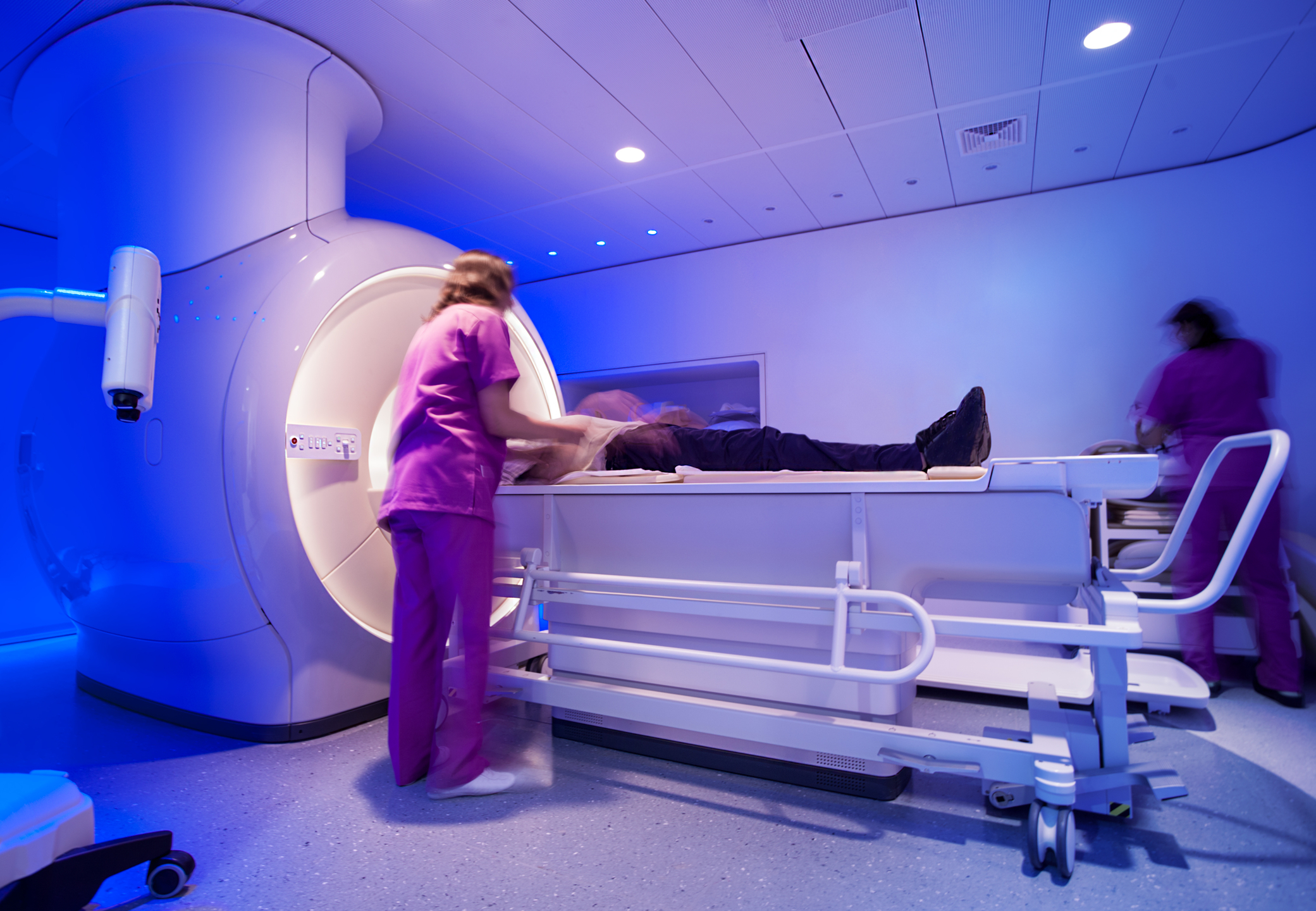
Explore the latest innovations in MRI equipment and how they enhance diagnostic imaging and improve patient care outcomes.
 By
By
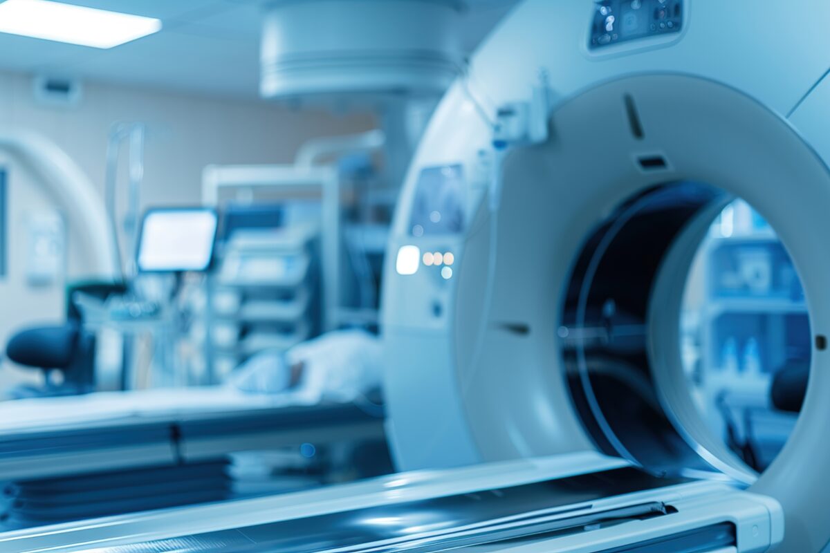
Understand the roles of medical scanners in modern medicine, highlighting their strengths and applications in healthcare diagnostics.
 By
By
Uncover the truth about castor oil and medical imaging. Learn why its benefits remain anecdotal and unverified by science.
 By
By
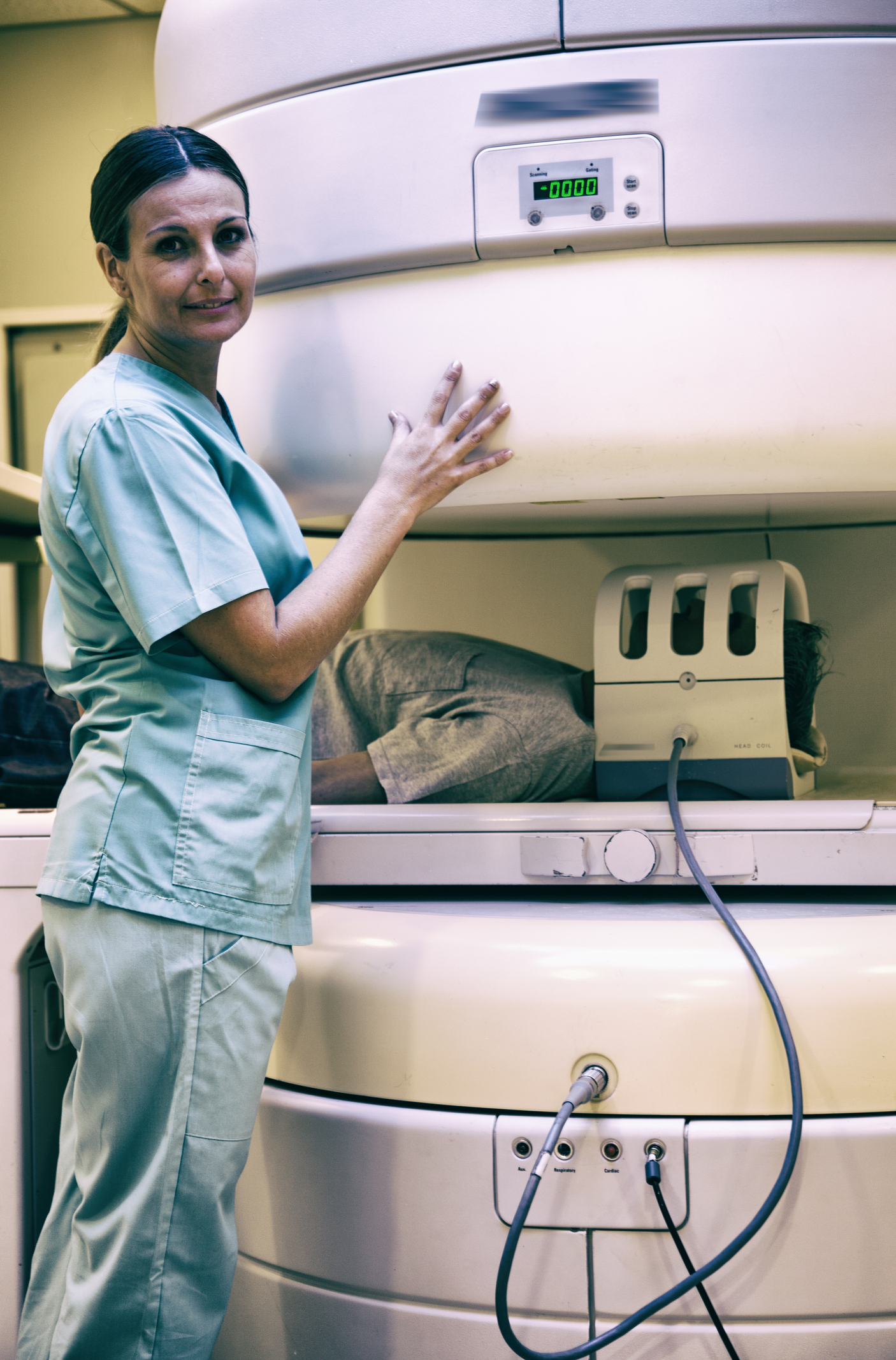
Uncover the technology behind Open MRI scanners and their role in advancing diagnostic imaging for patients and healthcare. Image for illustration only. People depicted are models.
 By
By
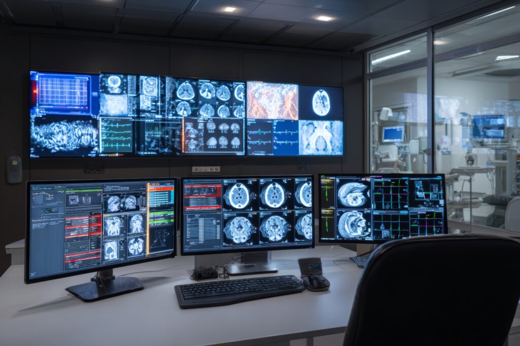
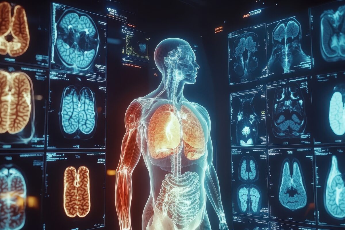
Discover how medical imaging for diagnosis has transformed healthcare with advanced technologies like MRI and CT scans.
 By
By
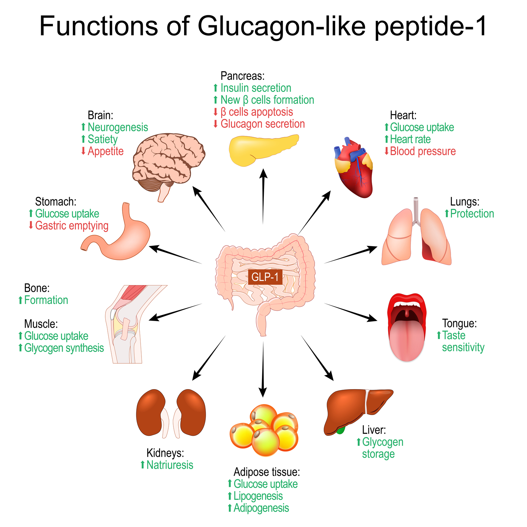
Discover how GLP-1 for weight loss functions as a powerful hormone, shaping our hunger signals and metabolic processes.
 By
By
Learn about the UK Biobank’s pioneering imaging study that has delivered unique insights from 100,000 participant scans.
 By
By
Uncover the vital role of Diagnostic Imaging Physics in modern medicine with insights into imaging technologies and patient safety.