SPECT Imaging
SPECT (Single Photon Emission Computed Tomography) is a nuclear imaging technique that allows clinicians and researchers to visualise the functional activity within the human body. SPECT imaging is particularly useful in neurology, cardiology, and oncology. It utilises radioisotopes that emit gamma radiation and a gamma camera to create detailed, three-dimensional images of the distribution of radioactivity within the body. As a result, SPECT provides valuable insights into the physiological processes, which can aid in the diagnosis, treatment planning, and monitoring of various diseases and conditions.
Principles of SPECT Imaging
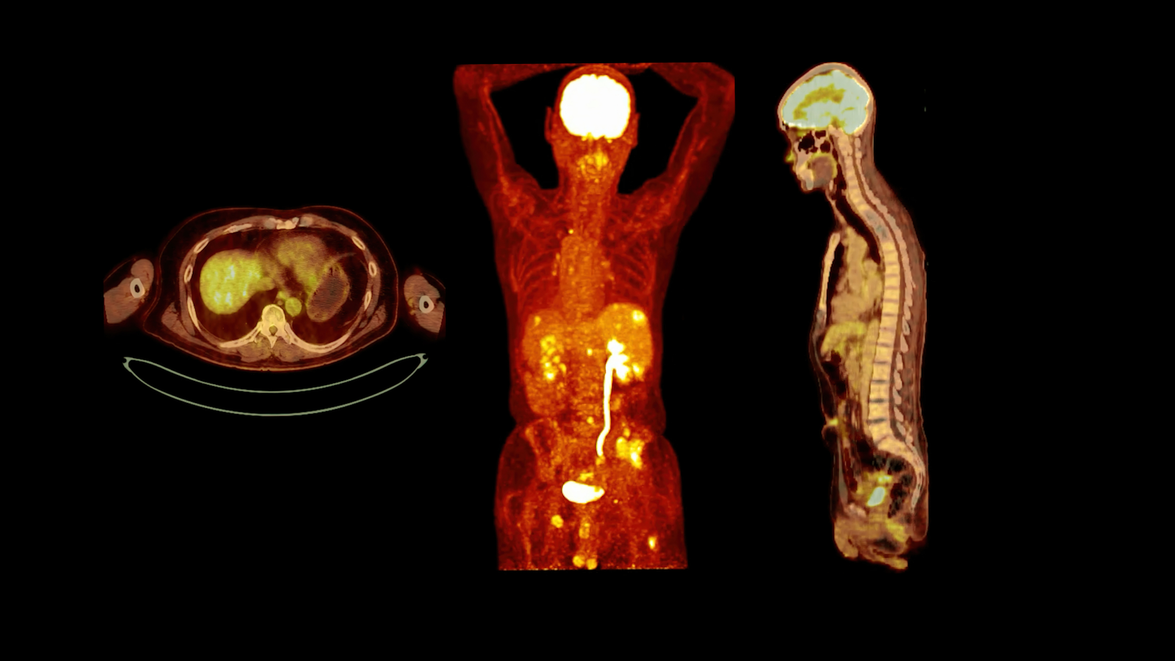
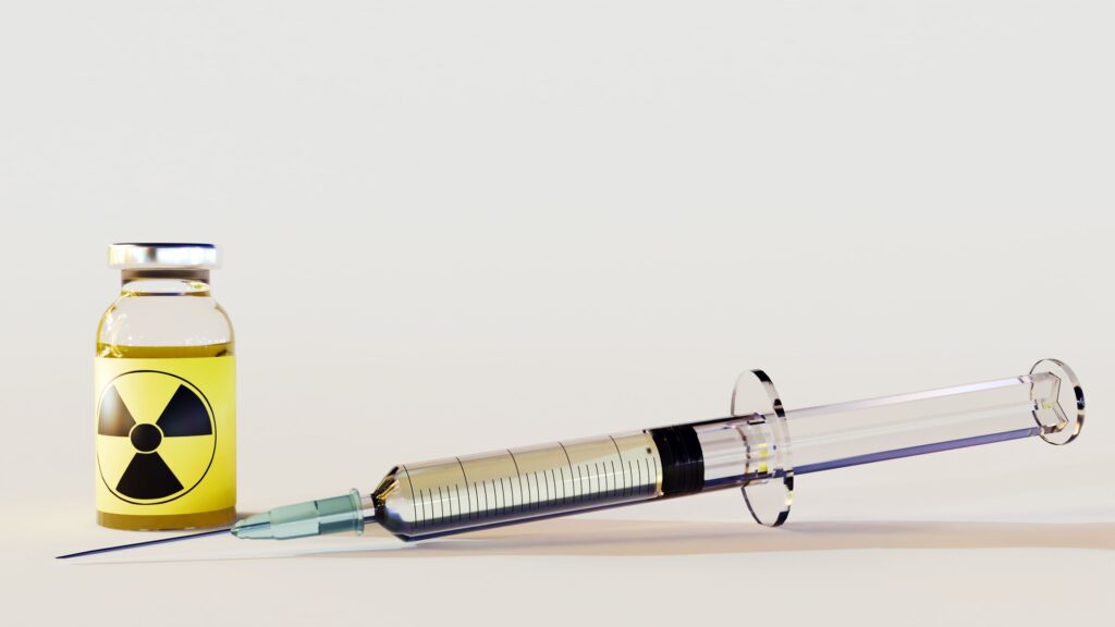
SPECT imaging relies on the use of radiopharmaceuticals, which are compounds containing a radioactive isotope that emit gamma rays. These radiopharmaceuticals are administered to the patient, typically through intravenous injection. Once inside the body, the radiopharmaceuticals accumulate in the target organ or tissue, depending on their chemical properties and affinity for specific biological processes. The distribution of radioactivity reflects the functional activity of the targeted area.
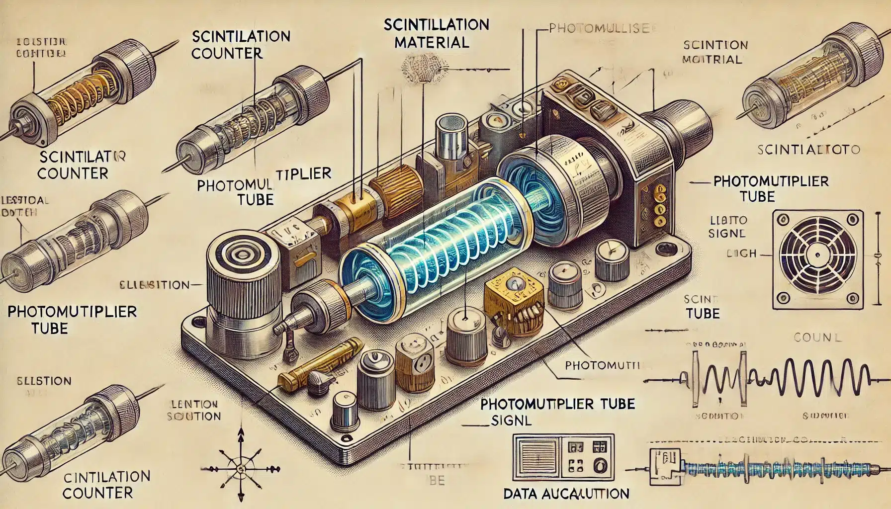
A gamma camera with a collimator detects and measures the emitted gamma radiation. The collimator filters and focuses the gamma rays onto a scintillation crystal, producing flashes of light proportional to the energy of the gamma rays. Photomultiplier tubes convert this light flashes into electrical signals that are processed and reconstructed into a three-dimensional image by a computer.
Applications of SPECT Imaging
- SPECT imaging has proven to be an invaluable tool for studying the brain’s functional activity. It is used to diagnose and monitor neurological disorders, such as Alzheimer’s disease, Parkinson’s disease, epilepsy, and stroke. By assessing regional cerebral blood flow, SPECT can provide insights into the underlying pathophysiology of these disorders.
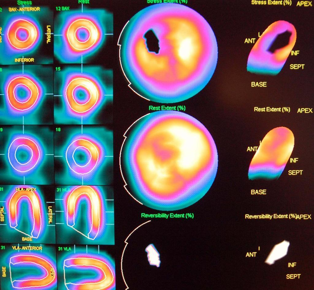
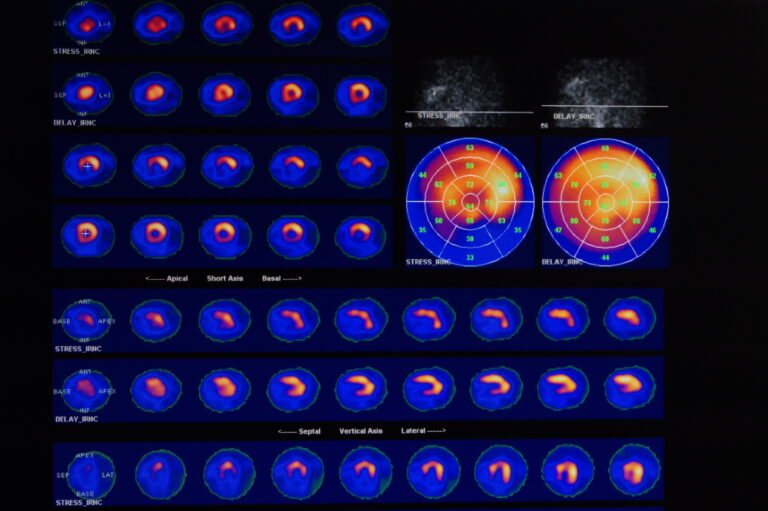
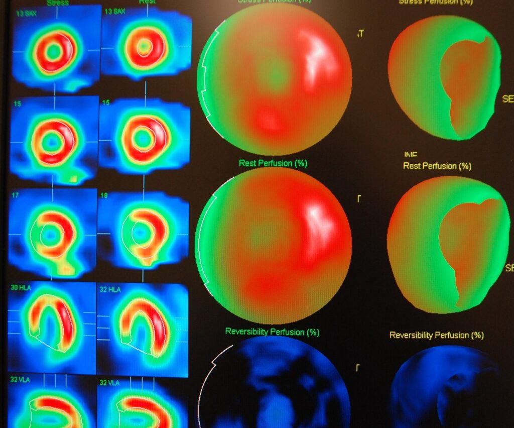
- SPECT imaging is used to assess myocardial perfusion, which reflects the blood flow to the heart muscle. This technique helps to identify areas of reduced blood flow, which may indicate coronary artery disease, and assess the heart muscle’s viability after a heart attack or before revascularisation procedures.
- SPECT imaging can identify and localise tumours, evaluate their metabolic activity, and monitor response to treatment. In addition, radiopharmaceuticals targeting specific cancer markers can be used to improve the sensitivity and specificity of SPECT for detecting malignancies.
home » SPECT imaging

 By
By