Why Emotional Support Is a Critical Part of Physical Injury Recovery
 By
By
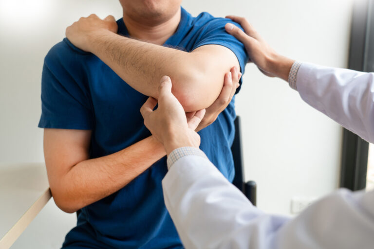
Emotional injury recovery requires patience, support, professional guidance, healthy routines, honest reflection, and time to rebuild confidence fully.
By
Emotional injury recovery requires patience, support, professional guidance, healthy routines, honest reflection, and time to rebuild confidence fully.
 By
By
Discover how dental health influences a child’s growth and confidence. Early care sets the stage for healthy smiles. Image for illustration only. People depicted are models.
 By
By
Discover how digital scanners in orthodontics are revolutionising treatment planning and diagnosis with unprecedented accuracy.
 By
By
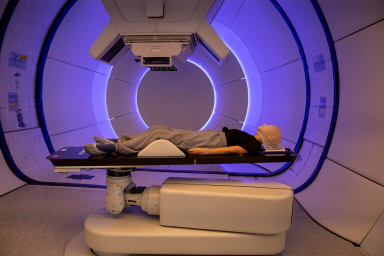
Learn about Modern Medical Imaging and Radiation Therapy and how advanced technology shapes the future of oncological treatment.
 By
By
Navigate the complexities of medical treatment cost in Germany. Find out what influences pricing for different healthcare procedures.
 By
By
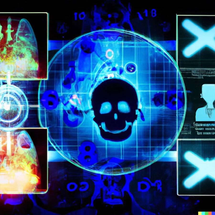
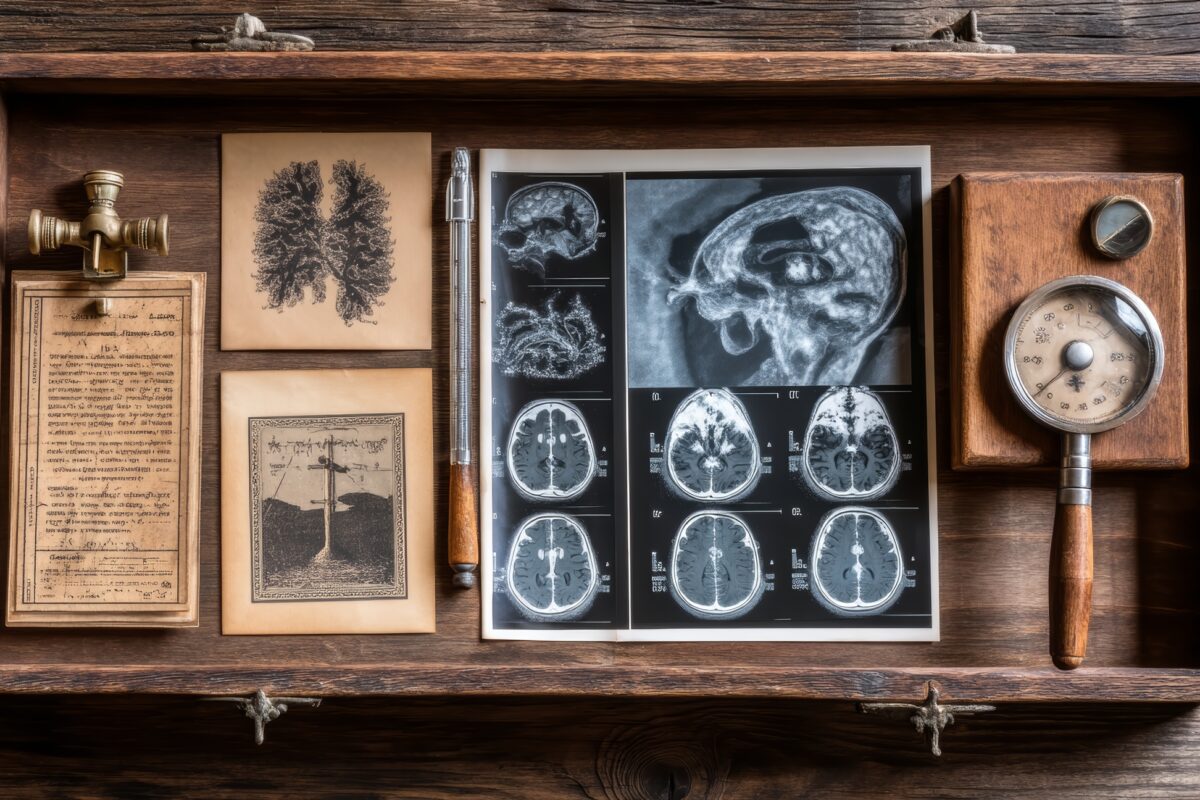
Explore the groundbreaking work of the pioneers of medical imaging and how they changed the landscape of modern medicine.
 By
By
Discover the impact of medical imaging in dentistry on modern practices, from enhanced diagnostics to revolutionary treatments.
 By
By
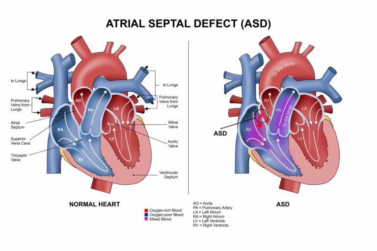
Understand the implications of atrial septal defect with insights on symptoms and necessary medical attention in adults.
 By
By
Learn key new nurse lessons to better prepare for your first job and anticipate the challenges ahead in nursing.
 By
By
Discover the fascinating types of medical imaging, from X-rays to advanced molecular techniques, shaping health care today.
 By
By
Learn about the truck accident health impacts that can persist for years. Discover the hidden challenges beyond initial injuries.
 By
By
Explore the potential of paediatric age estimation through deep learning and chest X-rays for accurate age assessment.
 By
By
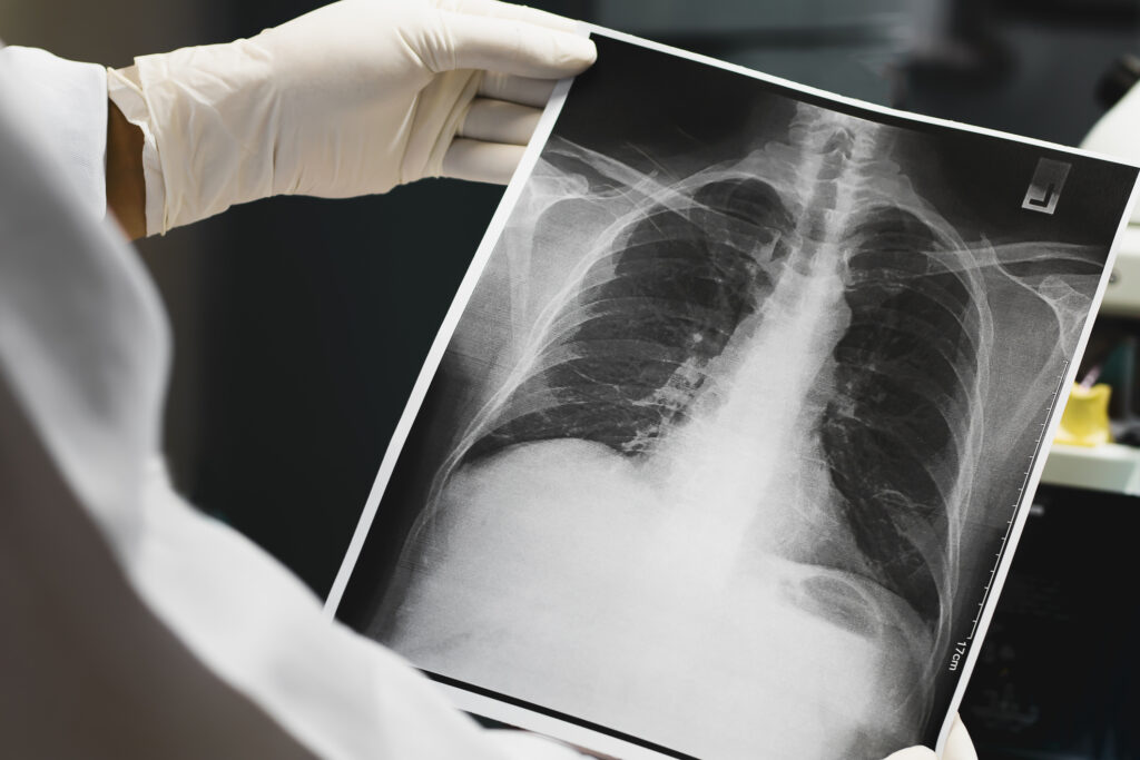
Discover the journey of X-rays in medicine through a real patient case and their importance in clinical diagnostics.
 By
By
Evaluate your expertise with the medical imaging quiz, focusing on imaging parameters, radiation safety, and practical case studies.
 By
By
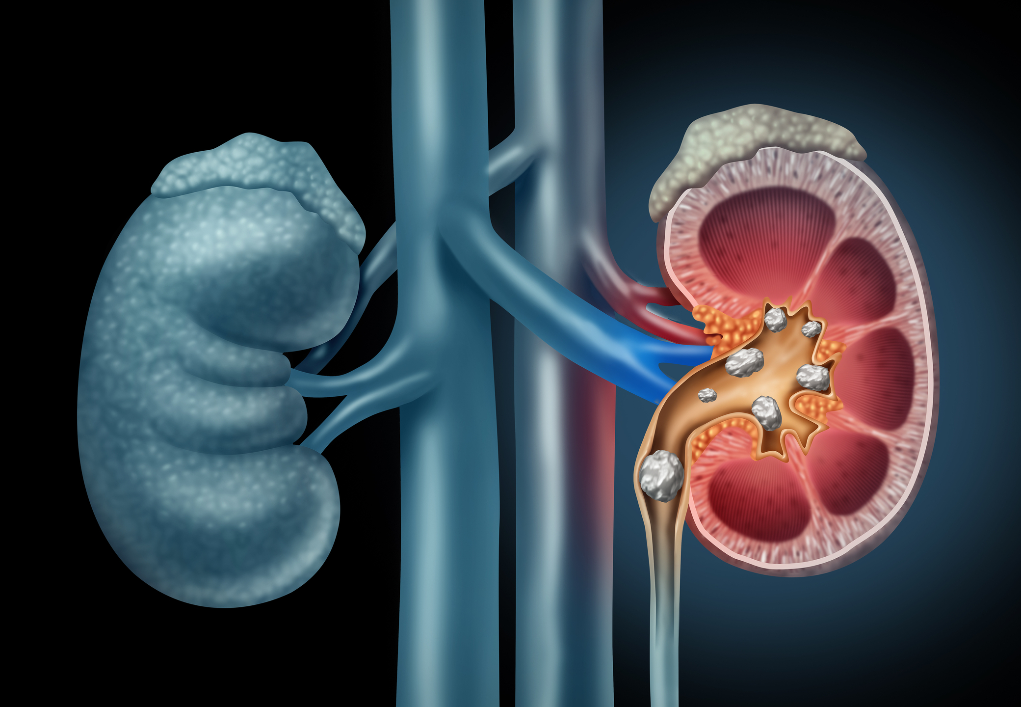
Discover advances in medical imaging in urolithiasis that improve the accuracy of renal colic diagnosis and treatment strategies.
 By
By
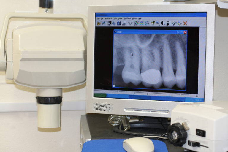
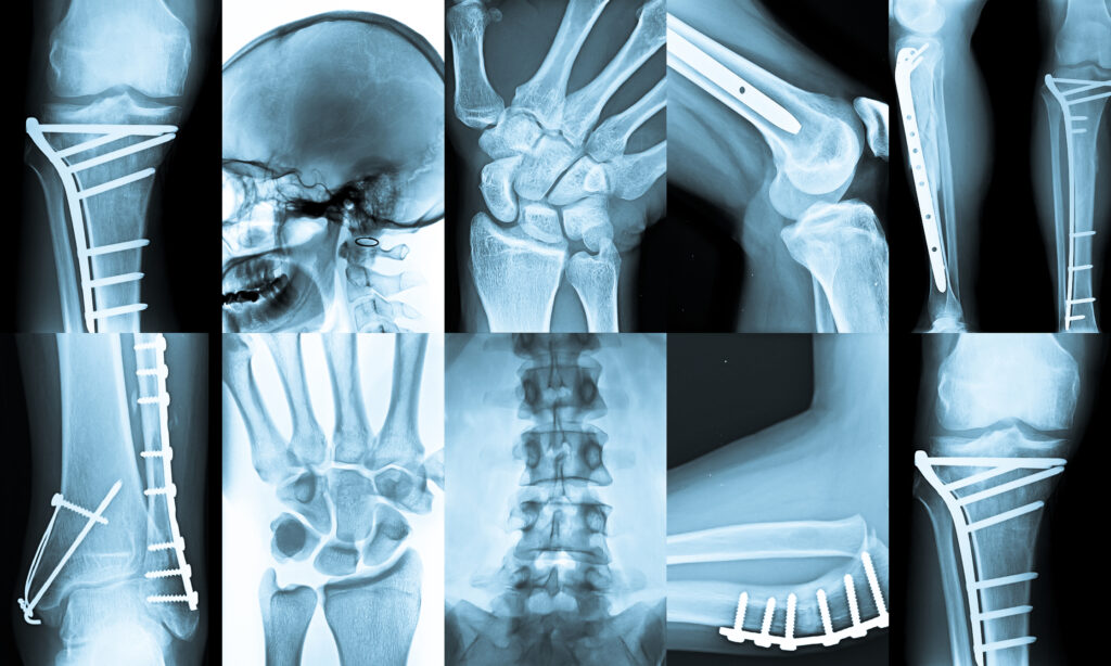
Understand the importance of medical X-rays in diagnostics and treatment, including their techniques and technological advancements.
 By
By
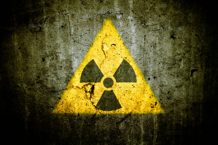
Explore what dark radiation is and its significance in medical diagnostics. Discover how it relates to X-ray technology.
 By
By
See a doctor after a car accident to catch delayed symptoms, begin treatment promptly, and strengthen any future injury claims. Image for illustration only. People depicted are models.
 By
By
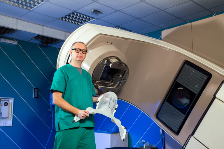
Megavoltage therapy units provide deep tissue radiation treatment, demanding accurate dosimetry and strict quality control procedures daily. Image for illustration only. Person depicted is a model.
 By
By
Find out about FLASH radiotherapy, the future of cancer care that delivers fast, effective radiation with fewer side effects.
 By
By
Low-speed crashes may look minor, but they often cause hidden spinal, ligament, and brain injuries over time.
 By
By
Explore the benefits of podiatry billing integration for managing patient loads and improving revenue in clinics.
 By
By
Undetected car accident injuries can worsen over time. Discover the warning signs and take action early for your health. Image for illustration only. Person depicted is a model.
 By
By
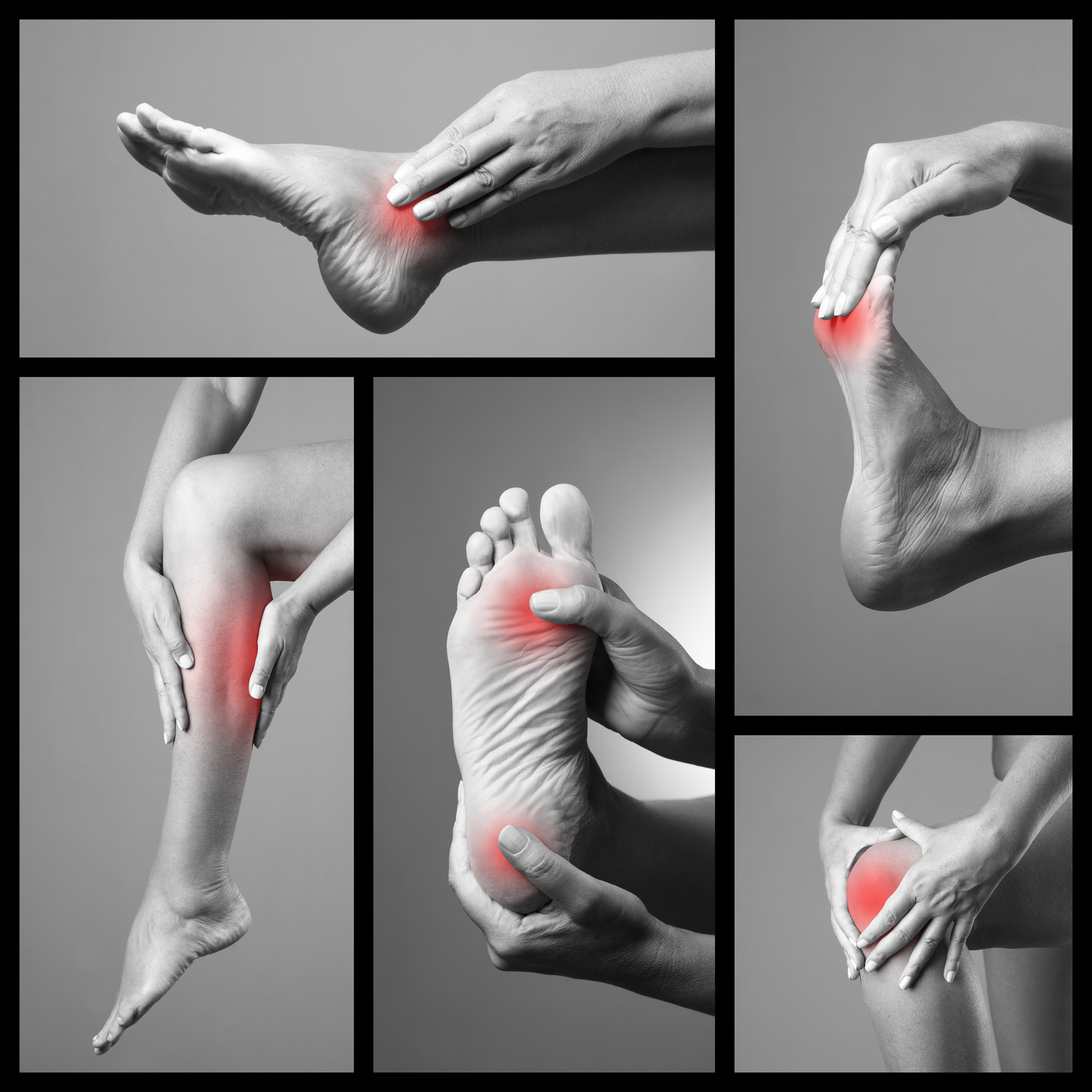
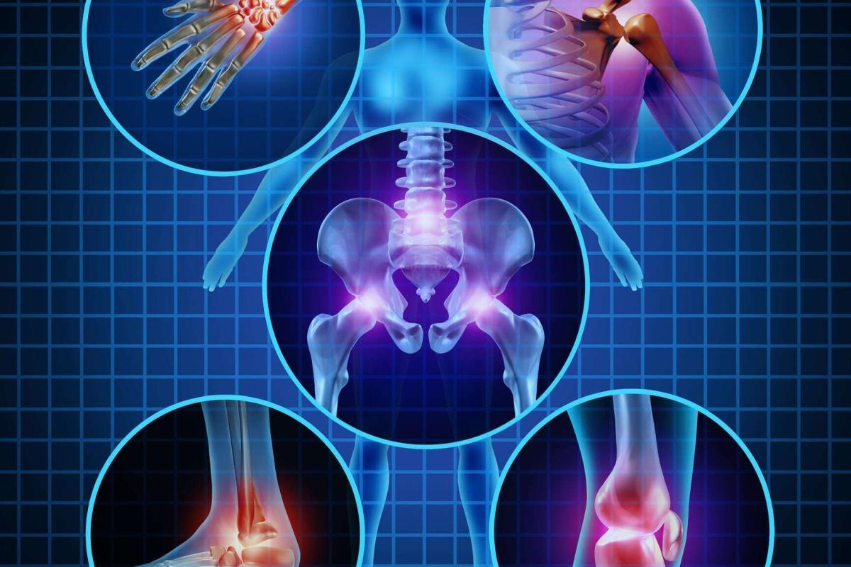
Knee health is essential for mobility, preventing pain, strengthening joints, improving flexibility, and maintaining an active, injury-free lifestyle daily.
 By
By
Medical imaging combines advanced technology, skilled professionals, and holiday warmth to support patients during Christmas emergencies.
 By
By
Sustainability in medical imaging highlights innovative practices, stakeholder roles, and strategies to reduce environmental impact effectively.
 By
By
Radiation dose management focuses on minimising exposure while maintaining diagnostic quality through advanced technologies and strategies.
 By
By
Magnetics in medicine enhance diagnostic precision, therapeutic targeting, and innovative treatments, transforming modern healthcare practices globally.
 By
By
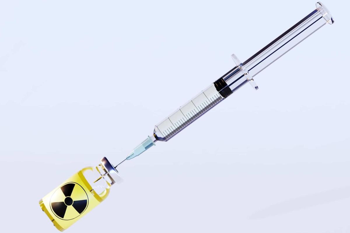
Nuclear Medicine Healthcare advances precision diagnosis, innovative therapies, and prioritises patient and professional safety.
 By
By
Sports medicine combines prevention, treatment, and rehabilitation to support athletes’ performance, health, and resilience.