How Medical Safety Gear Stops Hidden Germs
 By
By
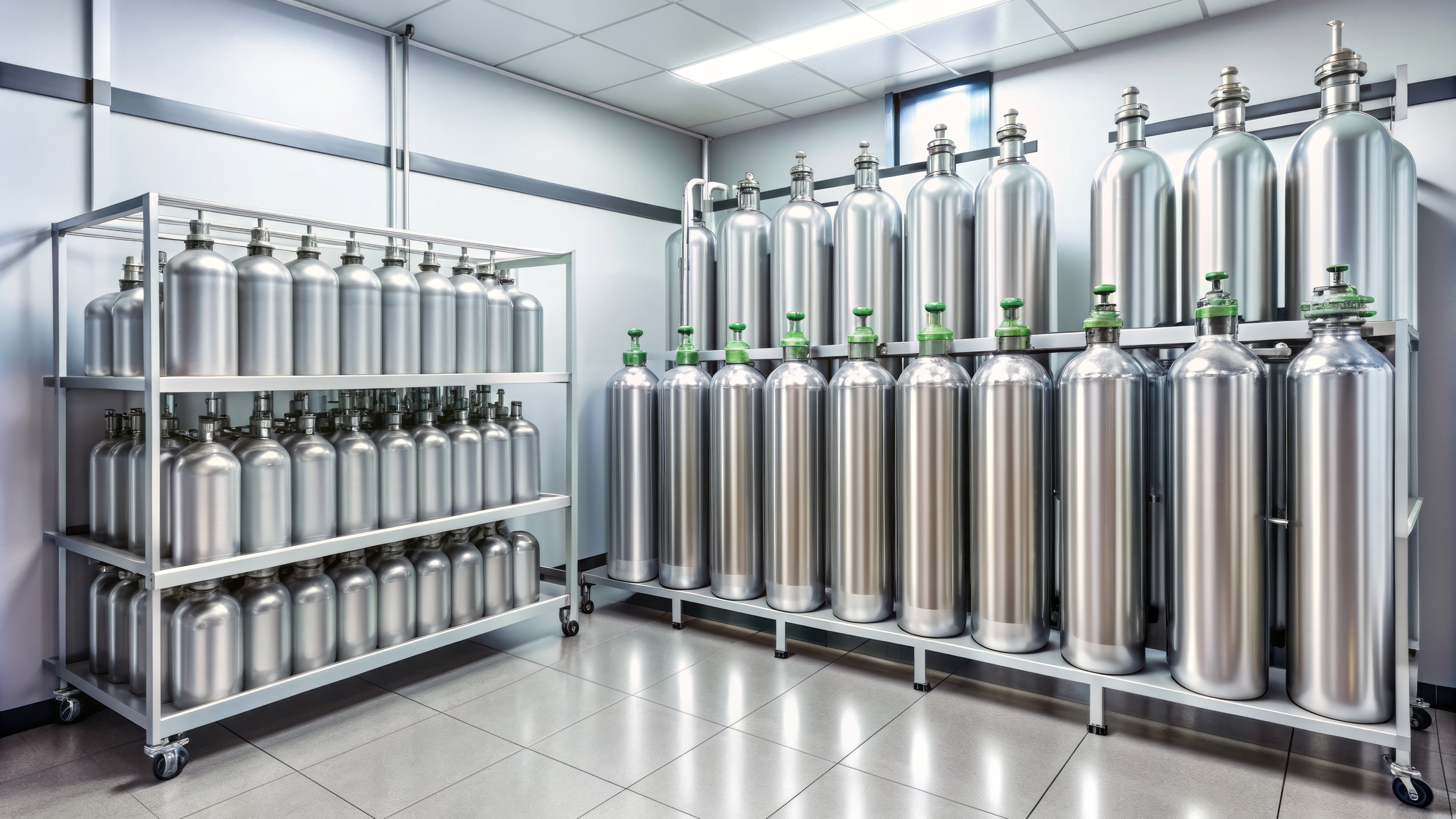
Learn how medical gas detection systems protect clinicians and patients from invisible threats in medical environments.
By
Learn how medical gas detection systems protect clinicians and patients from invisible threats in medical environments.
 By
By
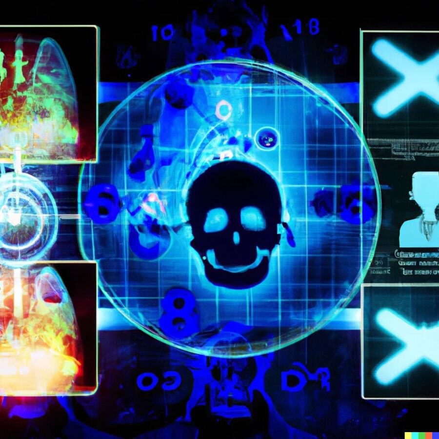
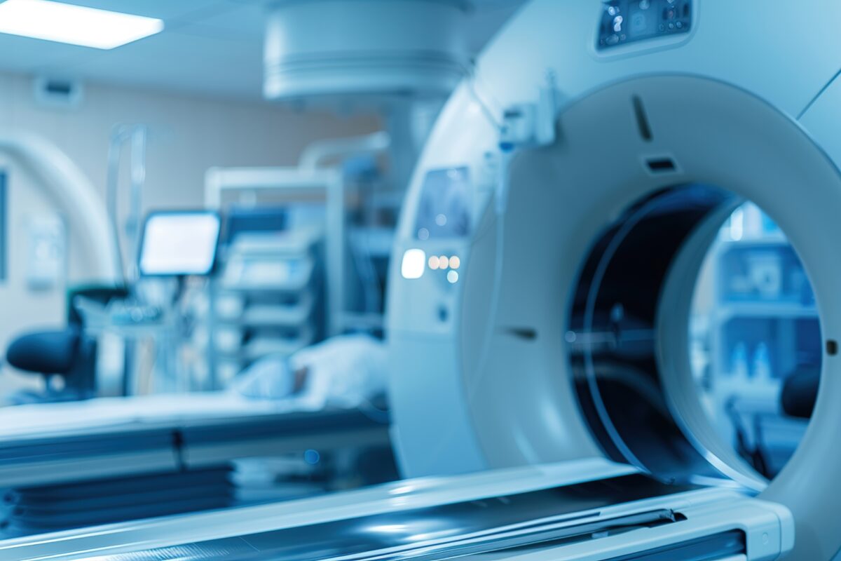
Learn about Modern Medical Imaging and Radiation Therapy and how advanced technology shapes the future of oncological treatment.
 By
By
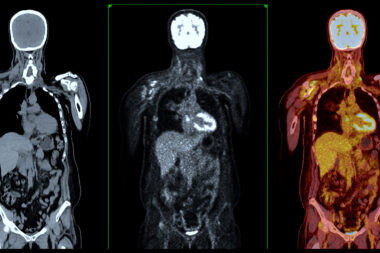
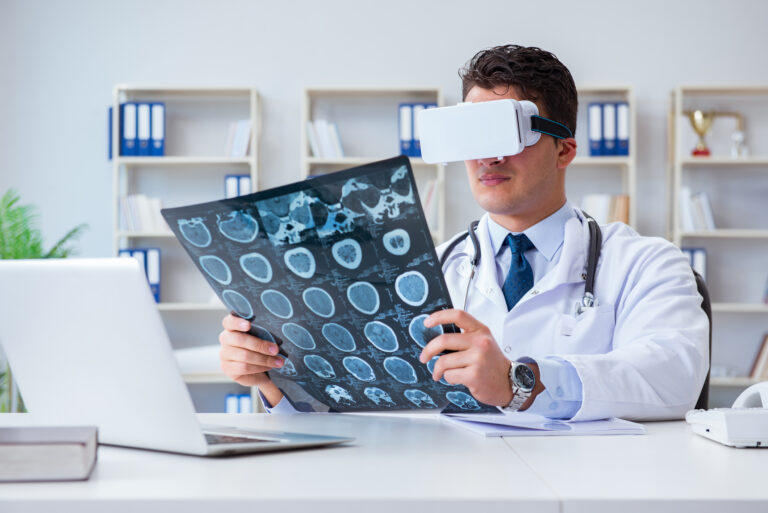
Stay informed on the innovations in medical imaging and healthcare, from artificial intelligence integration to precision medicine.
 By
By
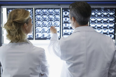
Discover the role of radiology and medical imaging in modern healthcare and how they enhance clinical decision-making. Image for illustration only. People depicted are models.
 By
By
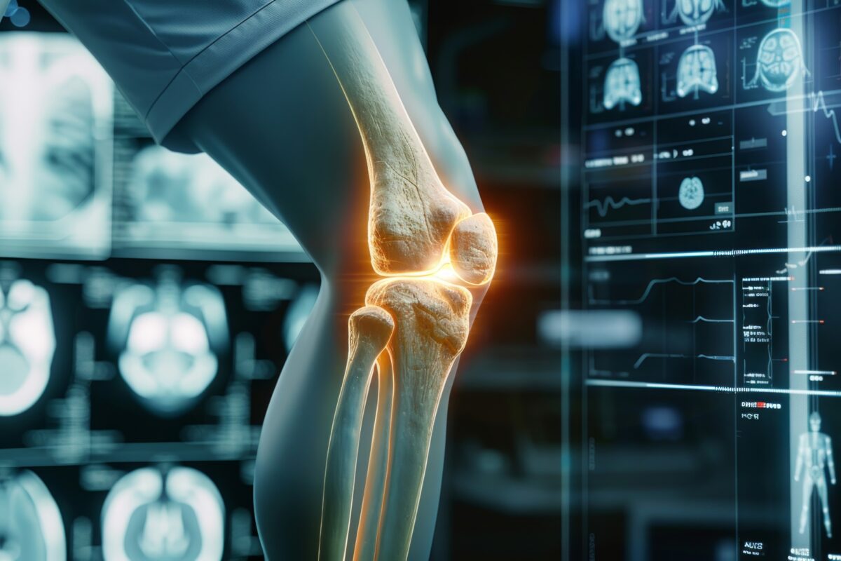
Discover the importance of musculoskeletal imaging diagnosis for accurate treatment of joint and back pain condition. Image for illustration only. People depicted are models.
 By
By
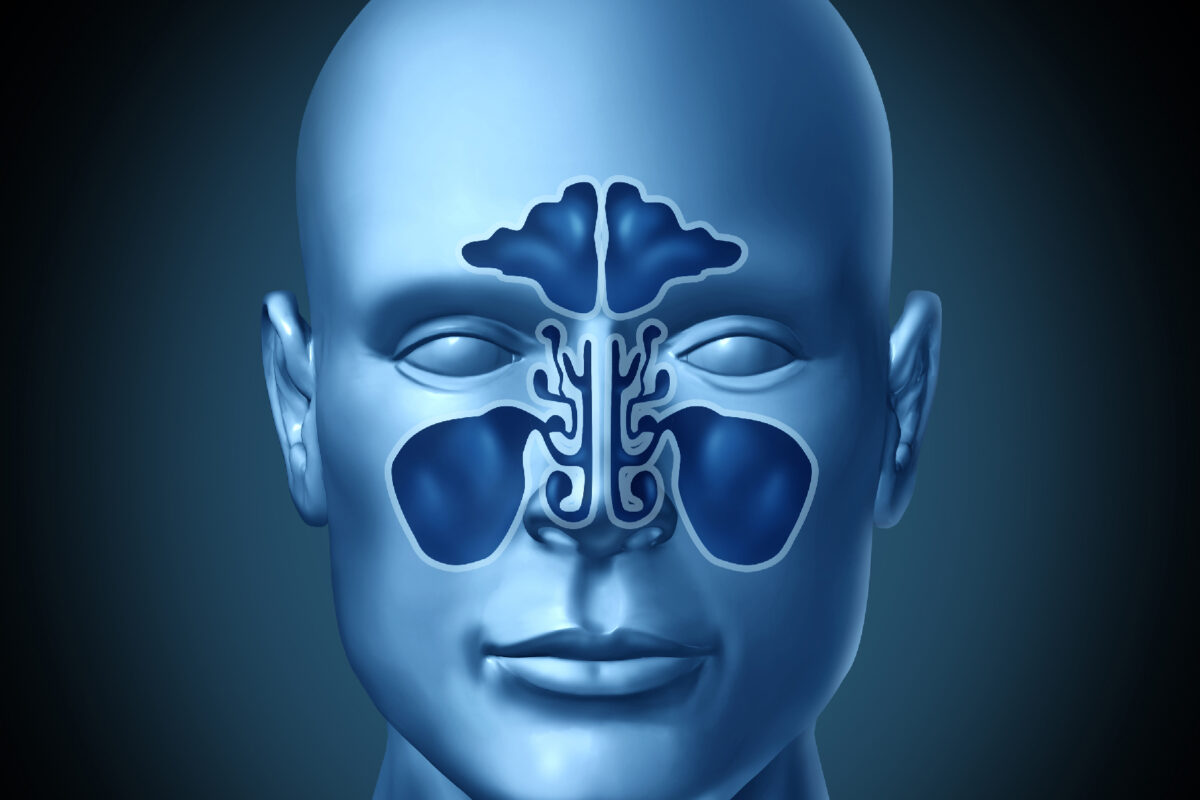
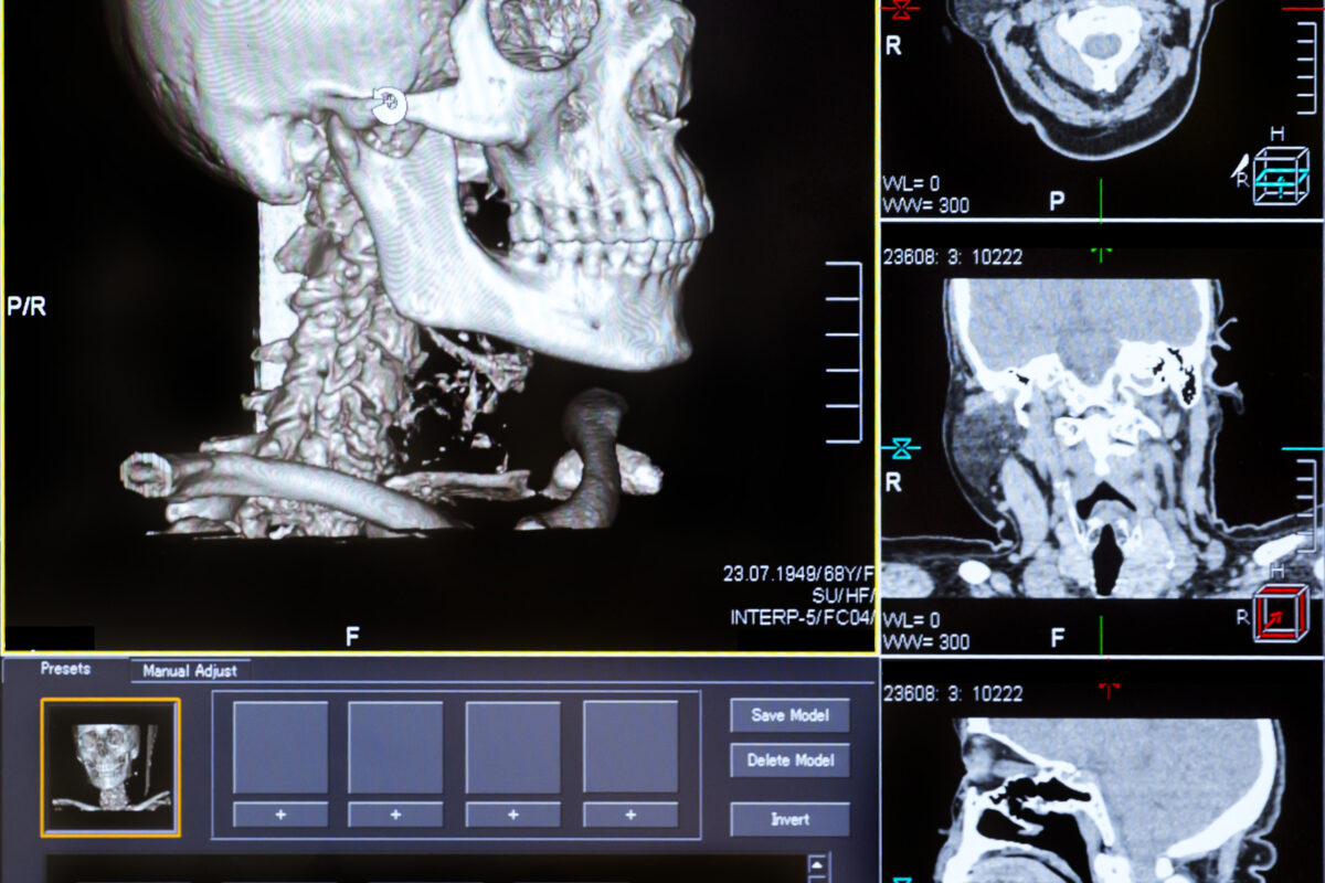
Discover how nasal and sinus imaging has evolved and its crucial role in diagnosing conditions like rhinosinusitis and tumours.
 By
By
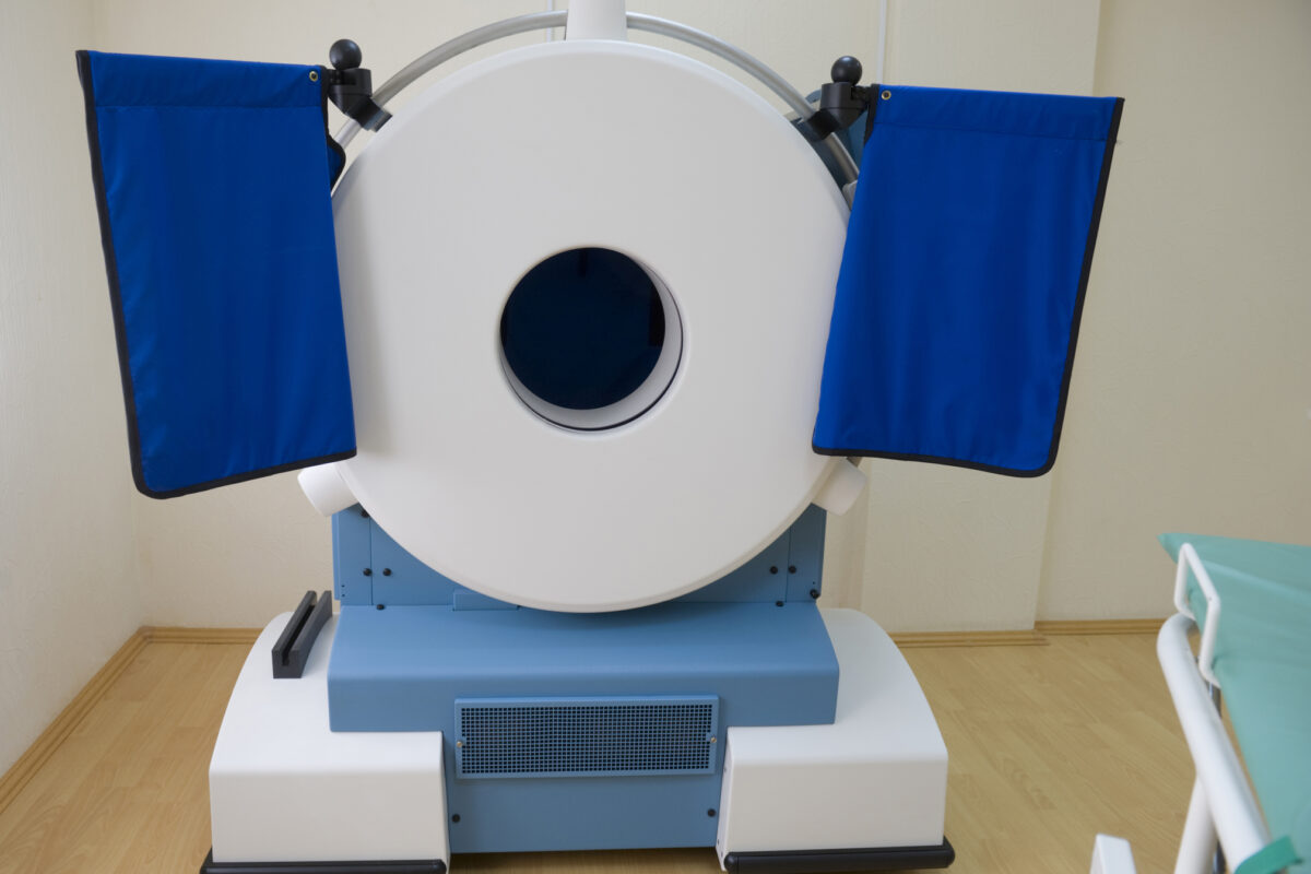
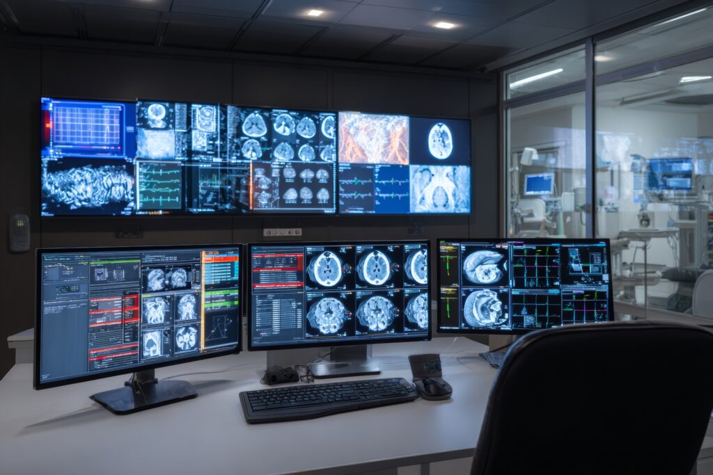
Mobile CT brain imaging is revolutionizing stroke care. Discover how this technology is used in various medical settings.
 By
By
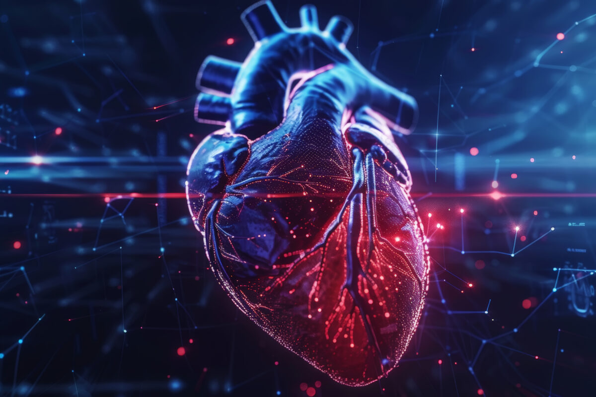
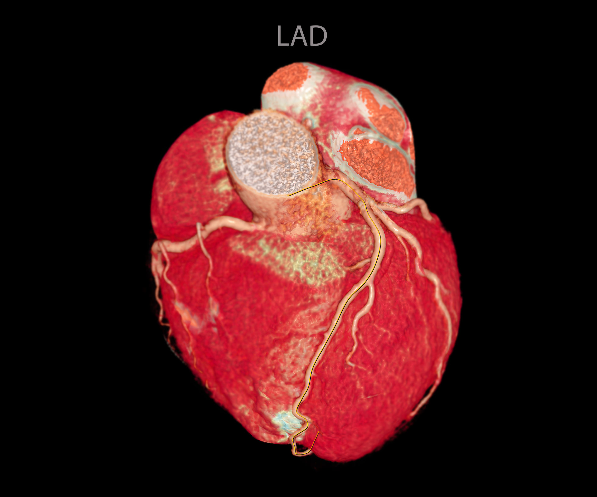
Discover how heart imaging is transformed by new technology, enhancing the diagnosis and treatment of cardiovascular disease.
 By
By
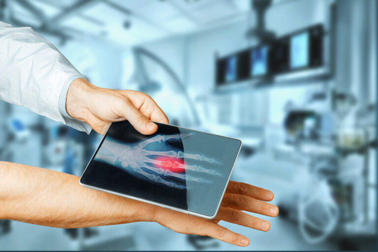
Learn about the latest developments in X-ray machines, including new technologies that redefine patient care and diagnostics.
 By
By
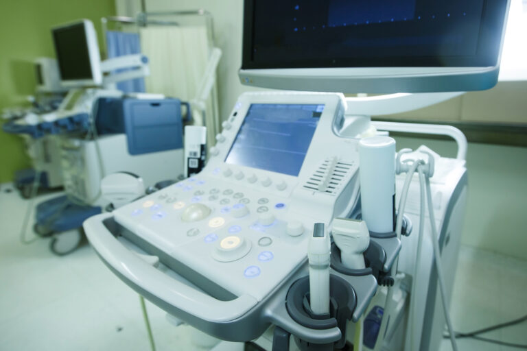
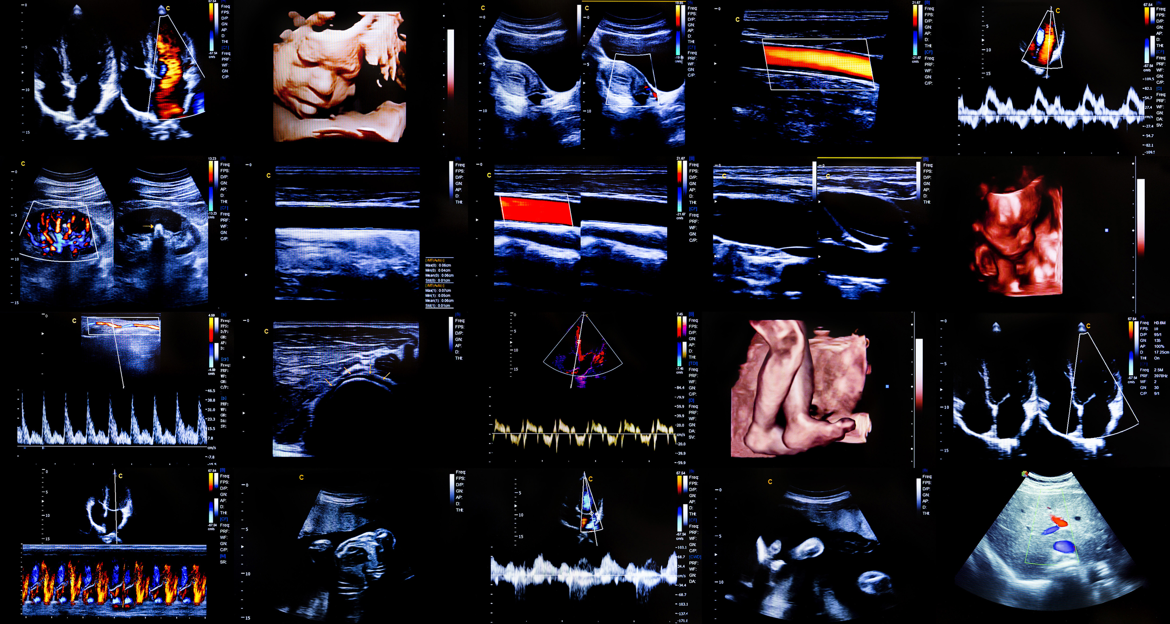
See how ultrasound technology is reshaping healthcare with automation, improving image quality and clinician efficiency.
 By
By
Uncover the latest innovations in photon-counting computed tomography and their impact on personalised healthcare and imaging.
 By
By
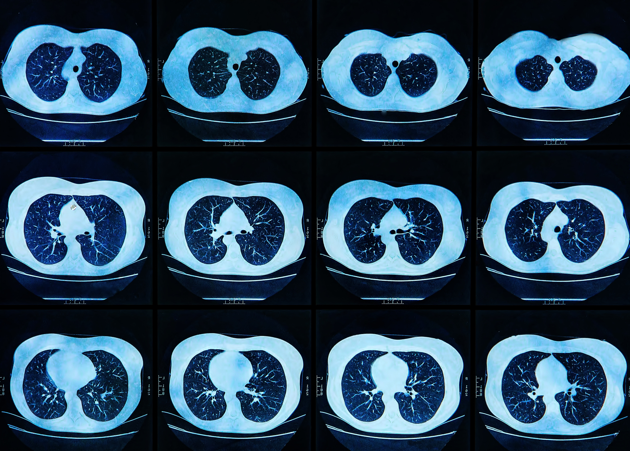
Discover how AI-powered low-field MRI is transforming lung imaging, enhancing accessibility and affordability for better healthcare.
 By
By
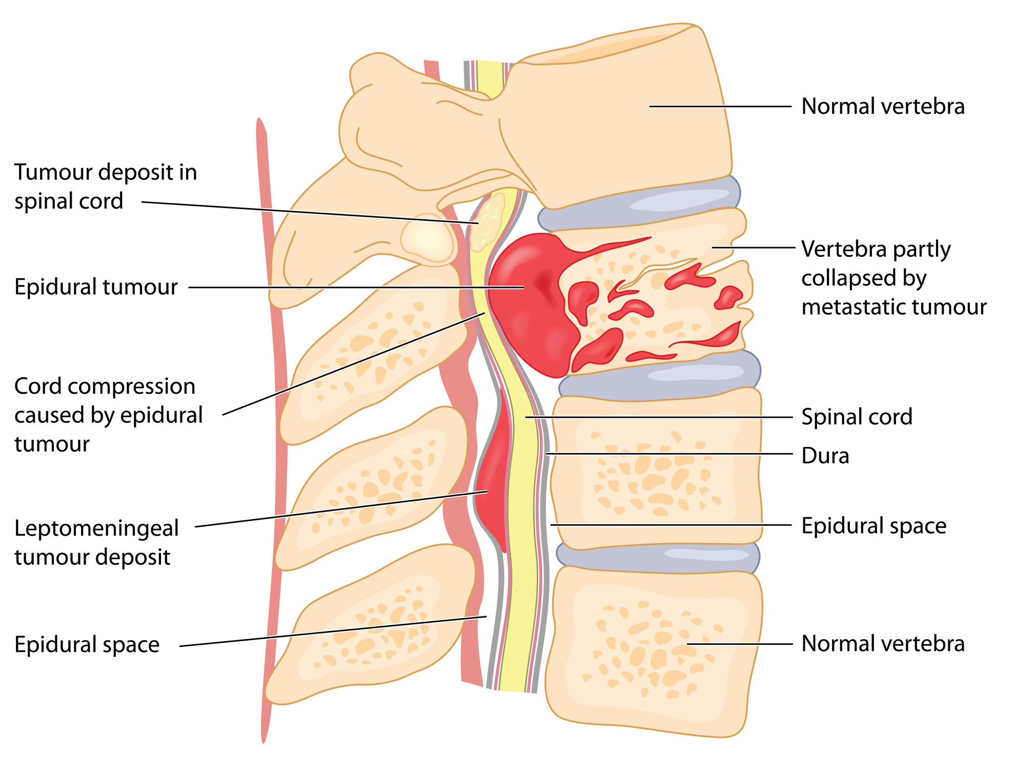
Discover the principles of MRI in spinal cord tumour diagnosis and how it helps identify various lesion types for improved care.
 By
By
Magnetic Resonance Angiography Aorta provides clear insights into vascular diseases, enhancing treatment planning and follow-up care.
 By
By
Uncover the benefits of advanced MRI sequences in multiple sclerosis, offering deeper insights into disease mechanisms and treatments. Image for illustration only. Person depicted is a model.
 By
By
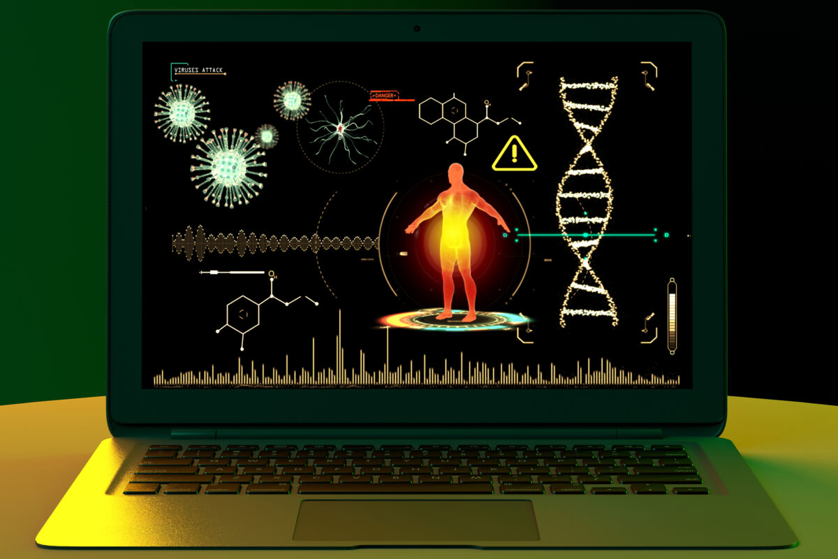
Discover how medical imaging for viral detection is revolutionizing diagnostics, offering insights into viral infections and treatments.
 By
By
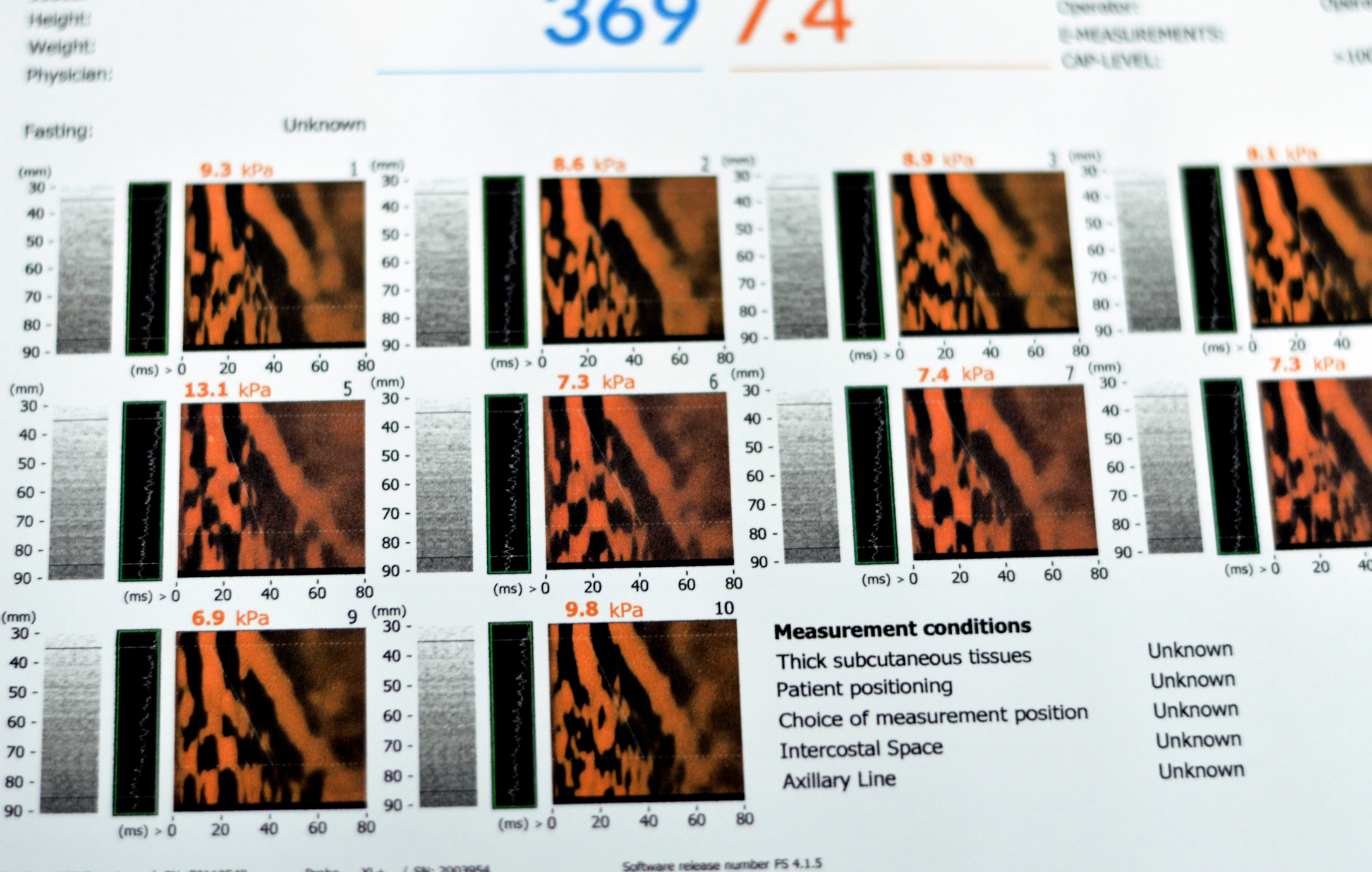
Understand the advantages of ultrasound elastography liver fibrosis over traditional biopsy in assessing chronic liver disease with precision.
 By
By
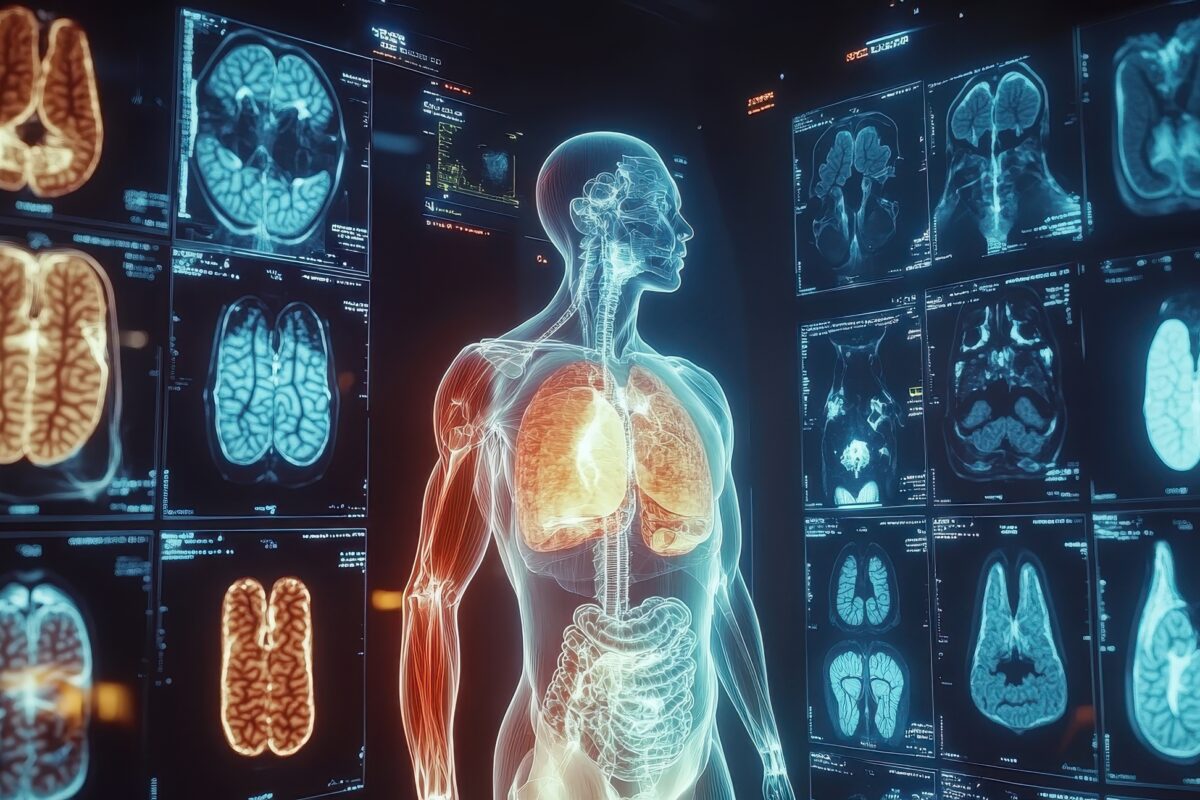
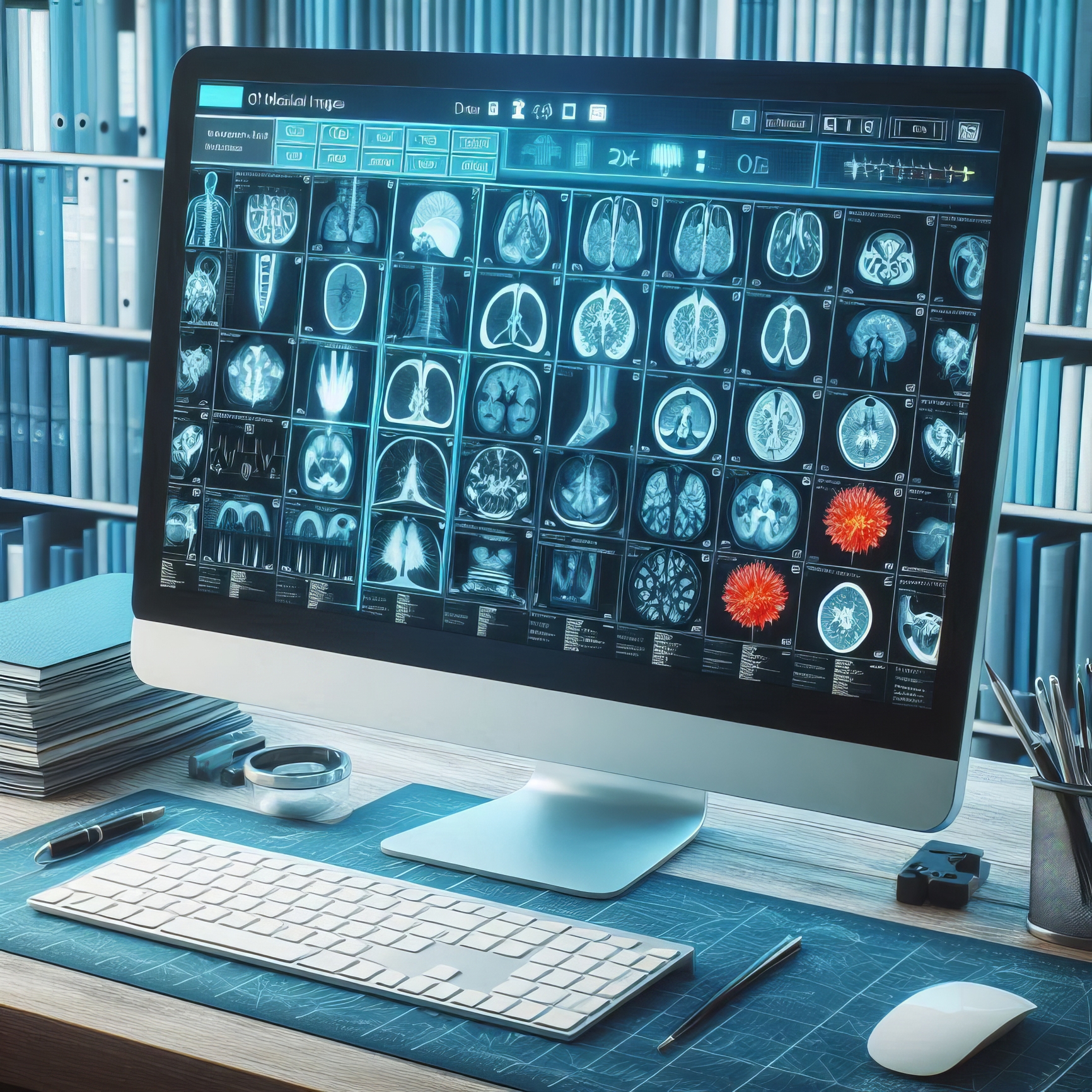
Discover the fascinating types of medical imaging, from X-rays to advanced molecular techniques, shaping health care today.
 By
By
Learn how microplate cell imaging allows for sensitive measurements and multiplexing in cellular assays for better research outcomes.
 By
By
Understand the roles of medical scanners in modern medicine, highlighting their strengths and applications in healthcare diagnostics.
 By
By
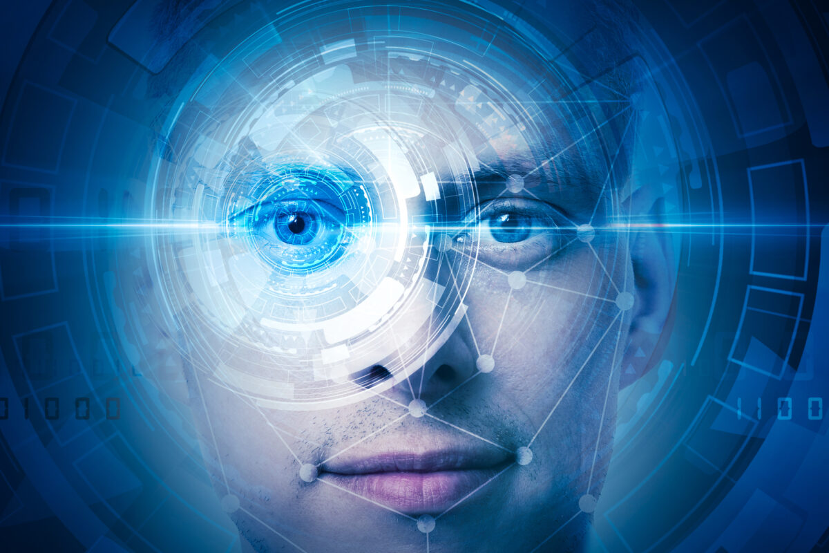
Learn about the role of facial confidence in interpersonal dynamics and how it affects our perception and communication. Image for illustration only. Person depicted is a model.
 By
By
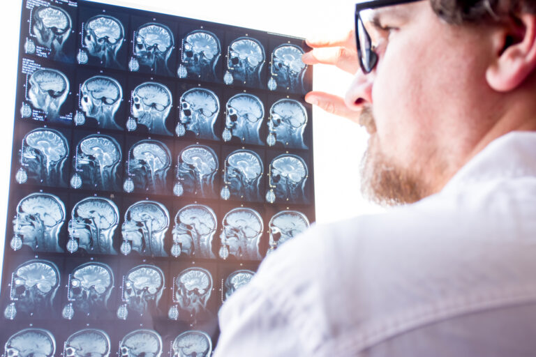
Discover how medical imaging for diagnosis has transformed healthcare with advanced technologies like MRI and CT scans.
 By
By
Uncover the vital role of Diagnostic Imaging Physics in modern medicine with insights into imaging technologies and patient safety.
 By
By
Uncover the principles of seeing with sound ultrasound and its potential future in non-invasive imaging and diagnostics.
 By
By
Explore the role of diagnostic X-rays and CT scans in modern medicine. Understand their applications and safety considerations.
 By
By
Diagnostic Imaging in Sports Medicine helps accurately diagnose injuries, guiding effective treatment and optimising athlete recovery timelines.
 By
By
New Year medical imaging breakthroughs highlight innovative technologies, transforming healthcare diagnostics and improving patient outcomes worldwide.
 By
By
Medical imaging combines advanced technology, skilled professionals, and holiday warmth to support patients during Christmas emergencies.
 By
By
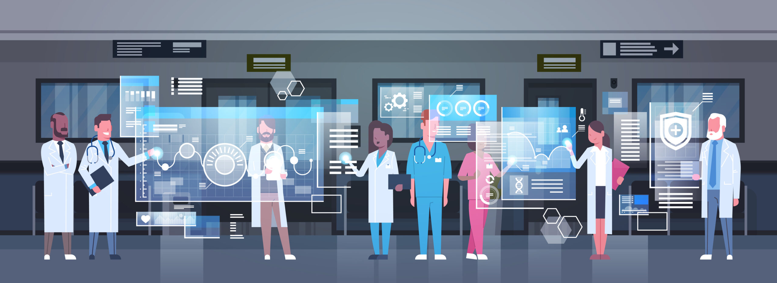
Open MedScience fosters global collaboration, accelerates innovation, promotes transparency, enhances accessibility, and transforms healthcare outcomes.
 By
By
Medical imaging 2025 integrates advanced AI, precision tools, seamless data systems, personalised diagnostics, and enhanced patient experiences worldwide. Image for illustration only. Person depicted is a model.