Total-Body PET and Beyond: The New Age of Molecular Imaging
 By
By
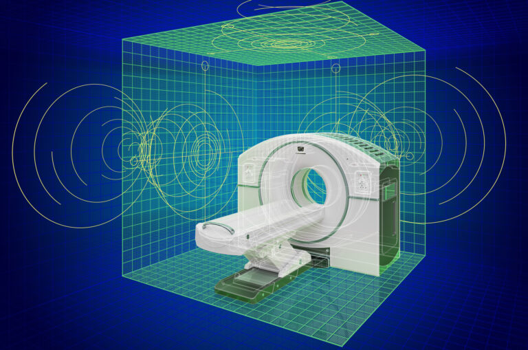
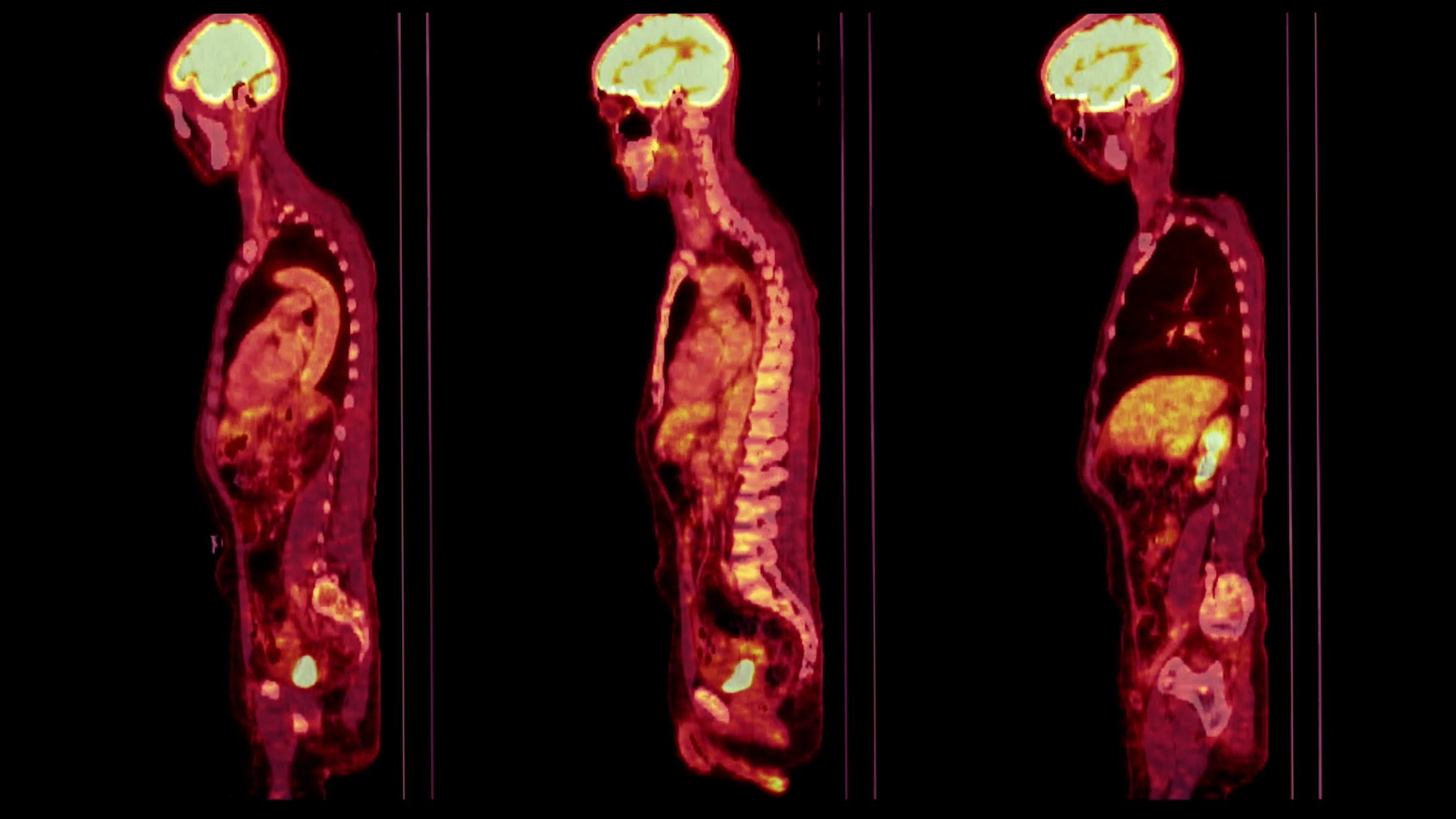
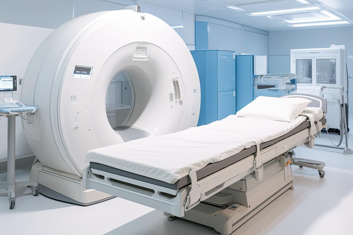
Explore total-body PET imaging and its advancements in sensitivity and clinical applications for better molecular insights.
By
Explore total-body PET imaging and its advancements in sensitivity and clinical applications for better molecular insights.
 By
By
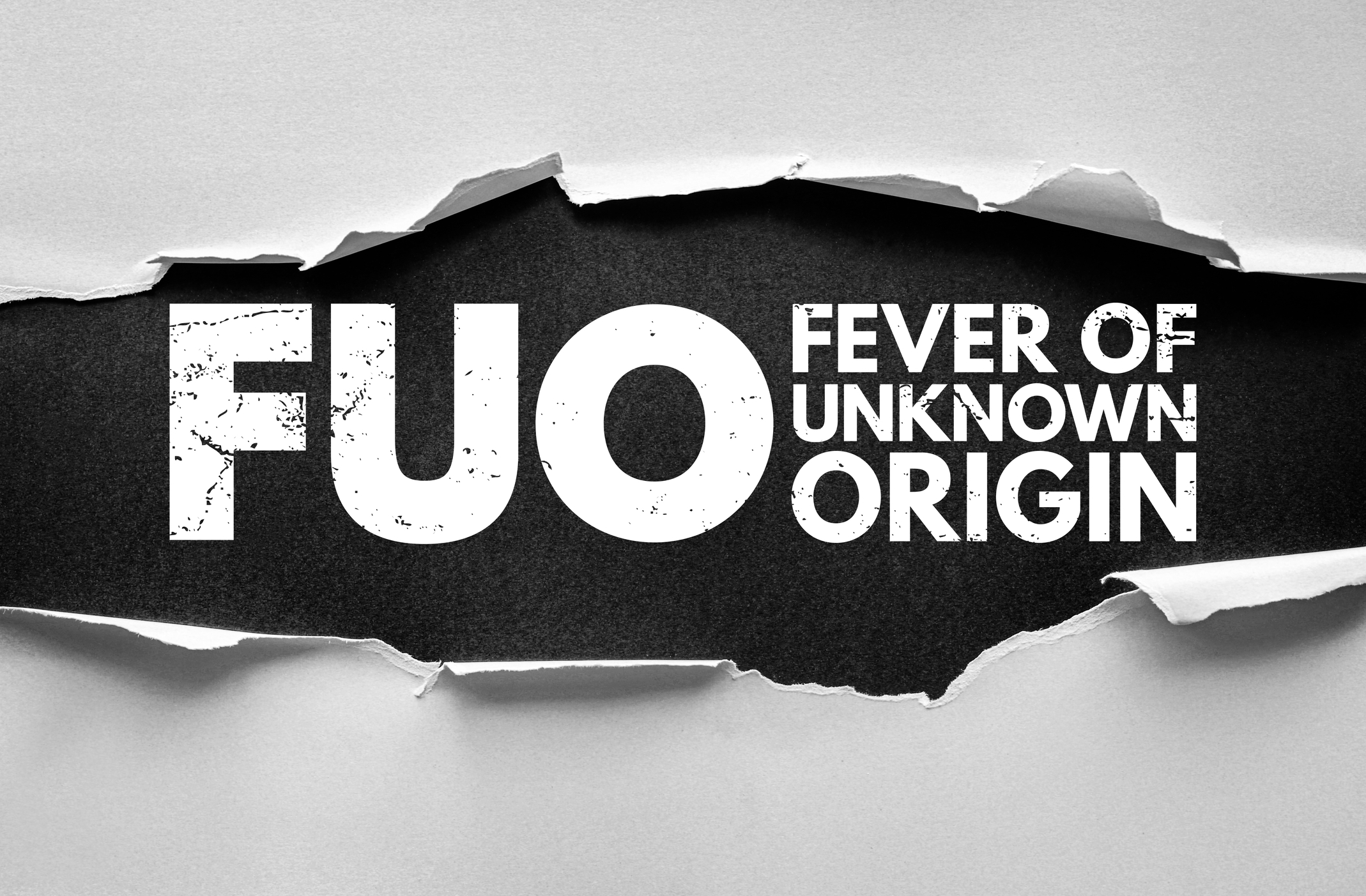
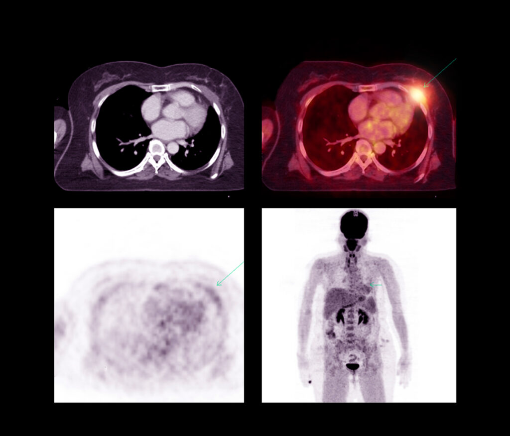
Uncover the benefits of using FDG PET-CT in fever of unknown origin and its impact on diagnostic accuracy and patient care.
 By
By
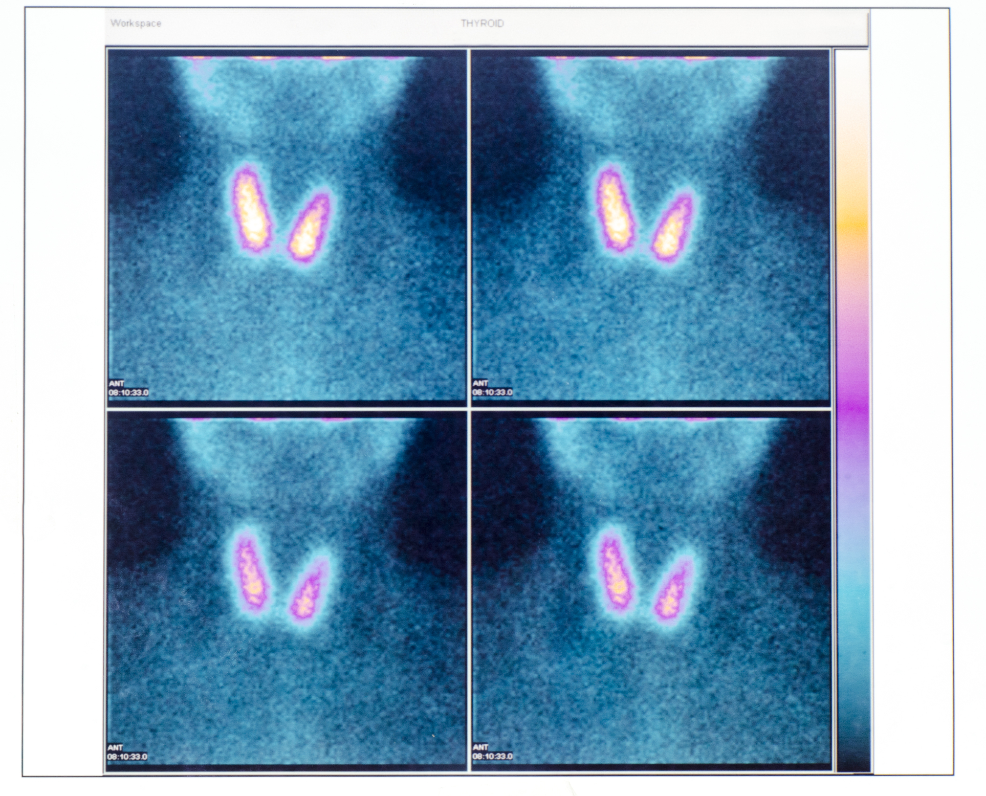
Uncover how PET/CT enhances thyroid cancer diagnosis. Explore its insights into tumours, staging, and personalised oncology.
 By
By
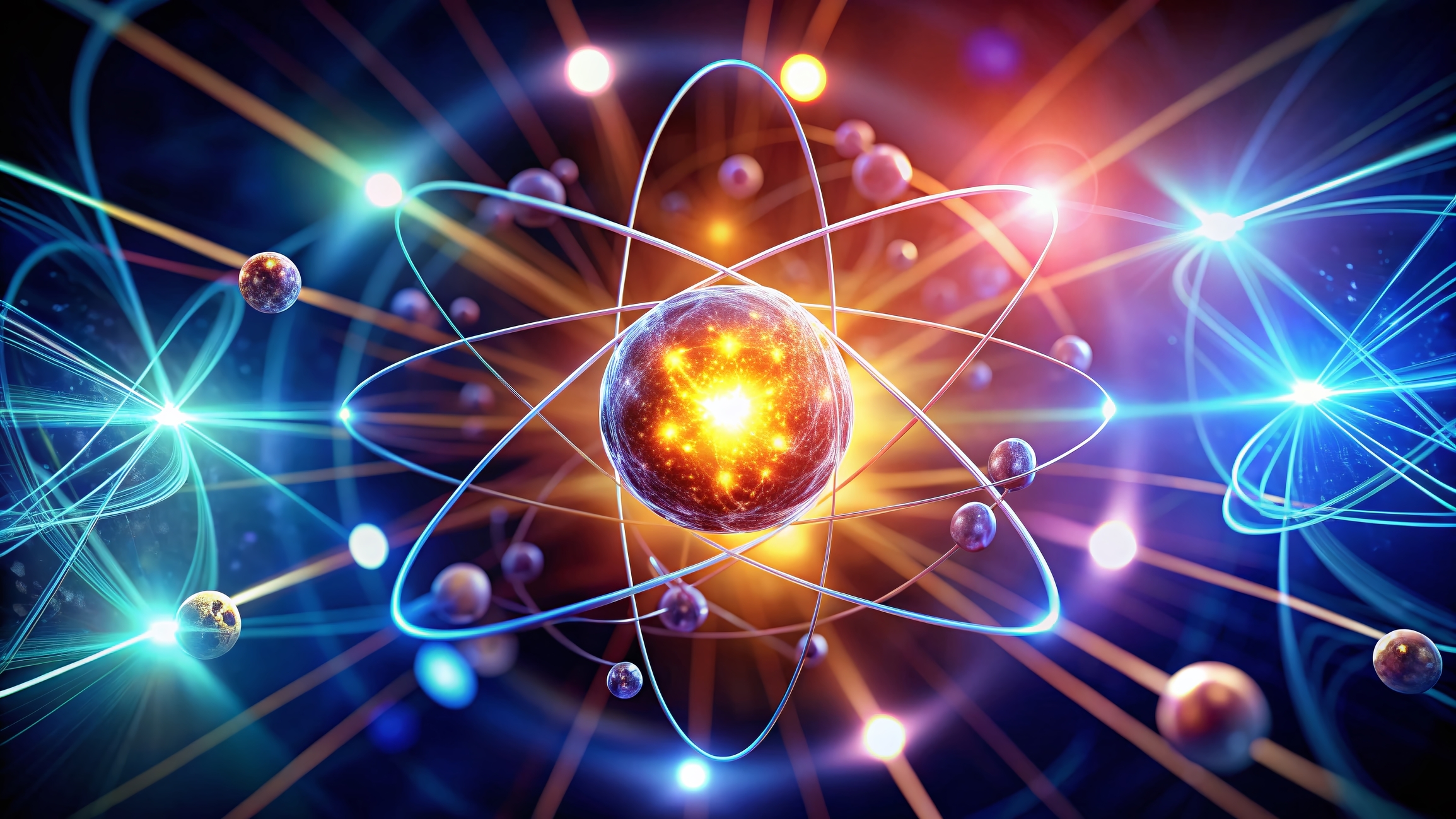
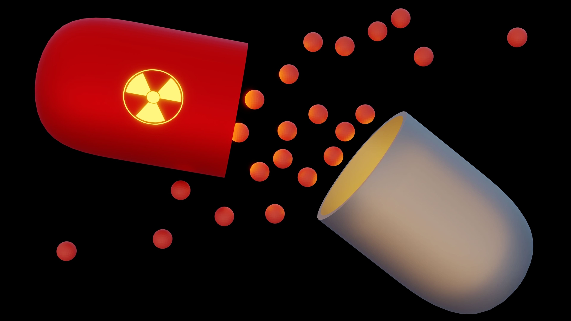
Discover how radiopharmaceuticals in medicine are transforming diagnostics and treatments with atomic precision for better health outcomes.
 By
By
Uncover the benefits of radioactive imaging. Understand PET, SPECT, and their role in advanced medical imaging and research.
 By
By
Learn how bioengineered contrast agents enhance imaging clarity and accuracy in nuclear medicine. Discover their unique benefits.
 By
By
Nuclear Medicine Healthcare advances precision diagnosis, innovative therapies, and prioritises patient and professional safety.
 By
By
Radiopharmaceuticals in diagnostics provide critical insights into disease processes, improving accuracy, patient care, and treatment outcomes significantly.
 By
By
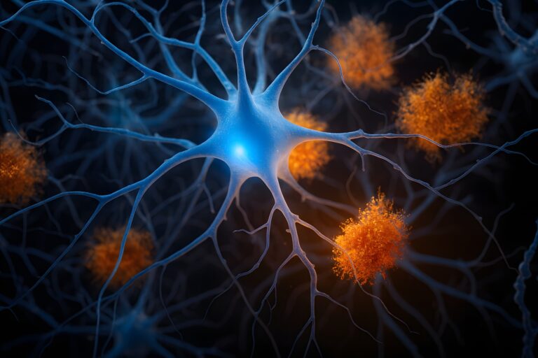
Pittsburgh Compound-B allows researchers to visualise amyloid plaques in the brain, aiding in Alzheimer’s disease diagnosis and study.
 By
By
Neuroimaging in nuclear medicine allows for detailed visualisation of brain activity, aiding in diagnosing complex neurological conditions.
 By
By
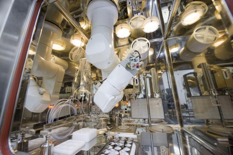
The synthesis of Fluorine-18 for advanced PET imaging requires precise cyclotron bombardment, purification, and automation technologies.
 By
By
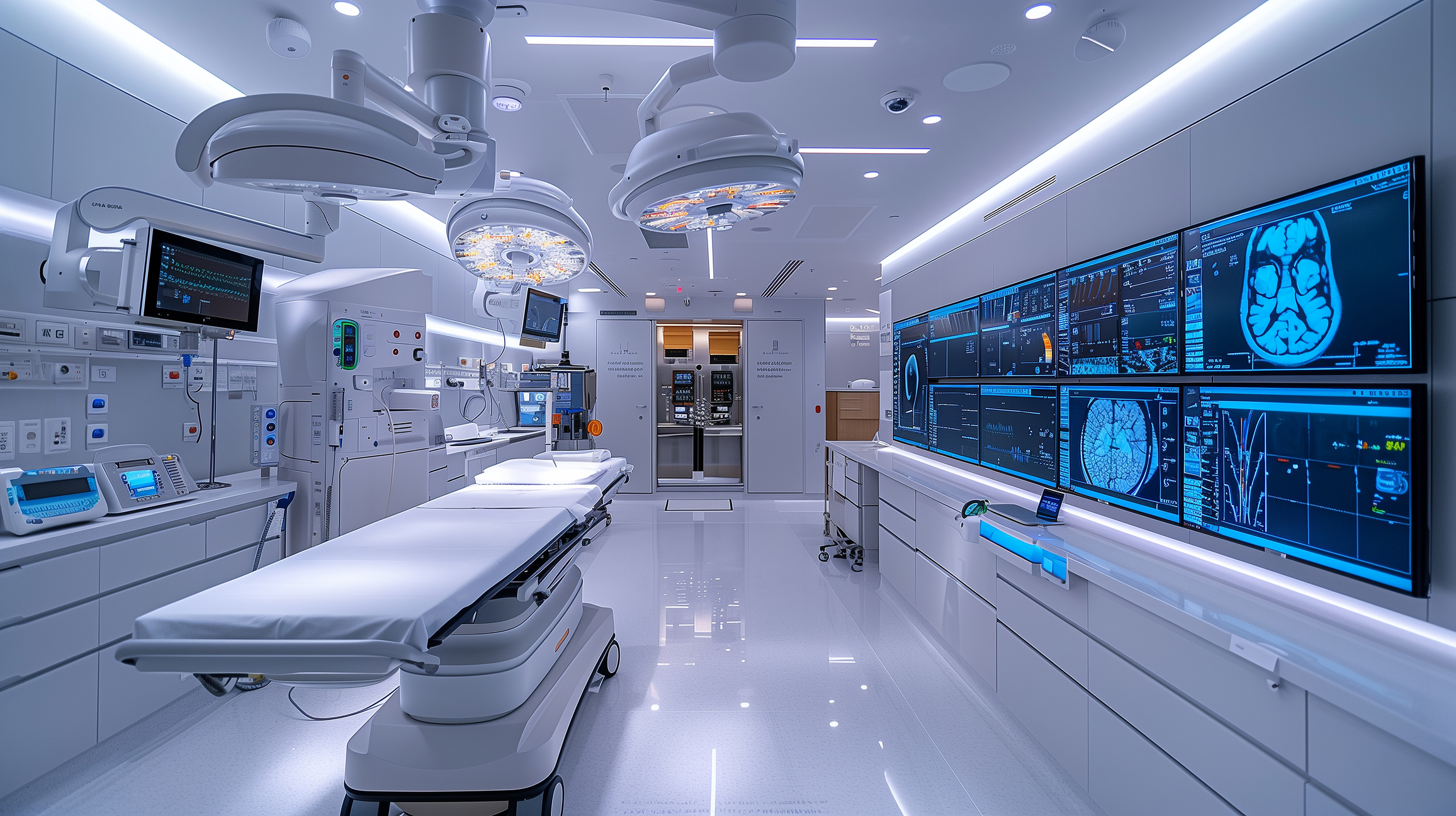
Positron Emission Tomography Imaging has advanced with cutting-edge technologies, enhancing diagnostic accuracy and expanding clinical applications dramatically.
 By
By
Nuclear medicine and the future of precise, personalised care in diagnosing and treating diseases.
 By
By
From Becquerel’s discovery in 1896 to modern medical applications, radionuclides have revolutionised our approach to science, medicine, and industry.
 By
By
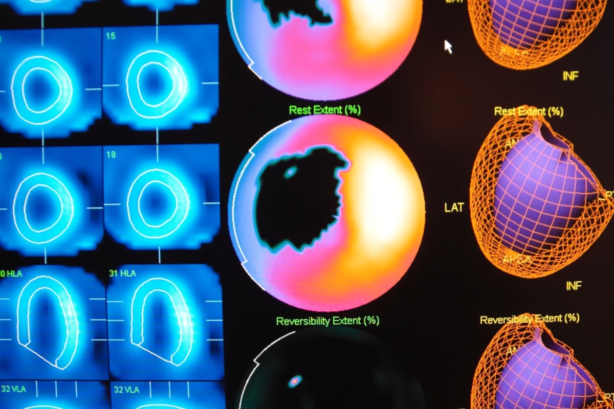
SPECT imaging provides valuable functional information, aiding clinicians in diagnosing, planning treatments, and monitoring progress.
 By
By
PET molecular imaging visualises cellular activity, aiding early diagnosis and tailored treatment for patients.
 By
By
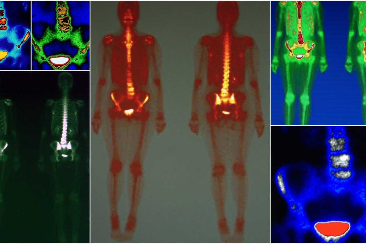
Bone imaging is an essential diagnostic tool for detecting bone diseases, injuries, and disorders.
 By
By
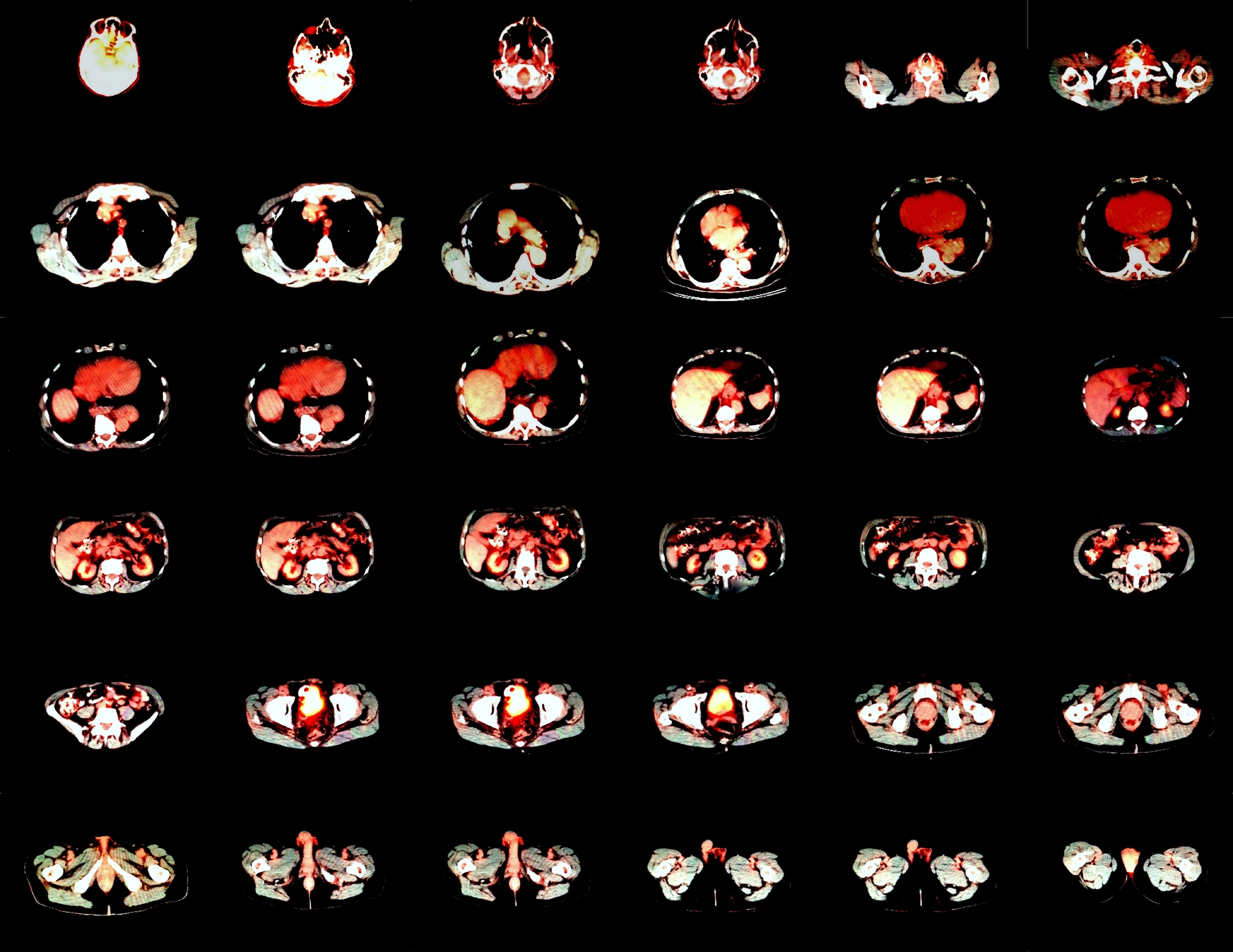
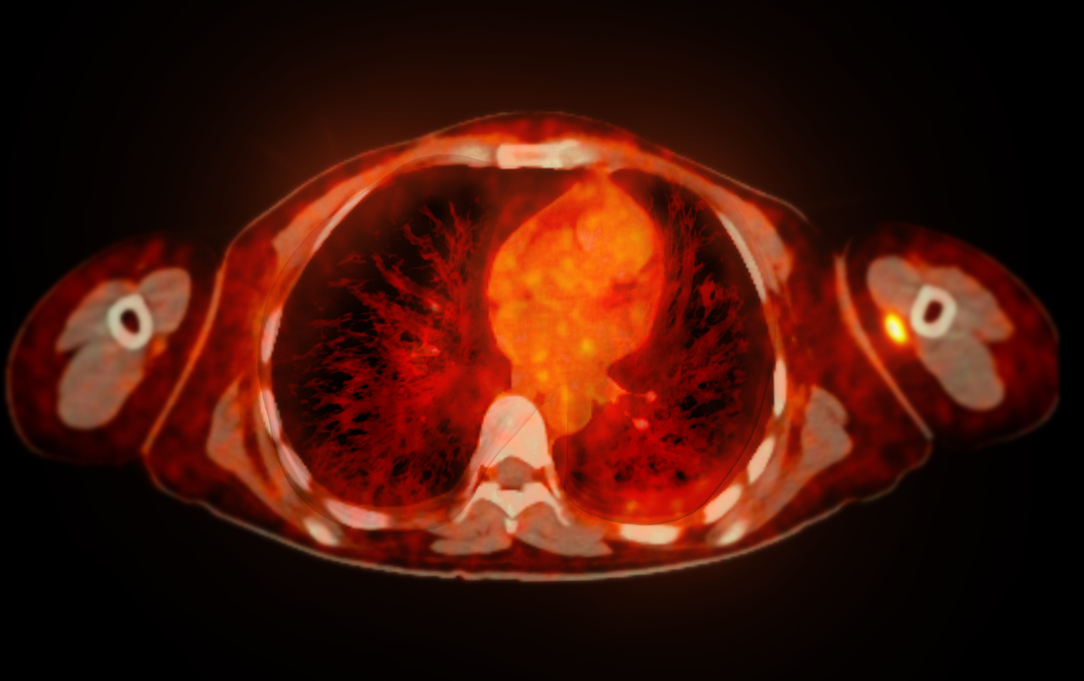
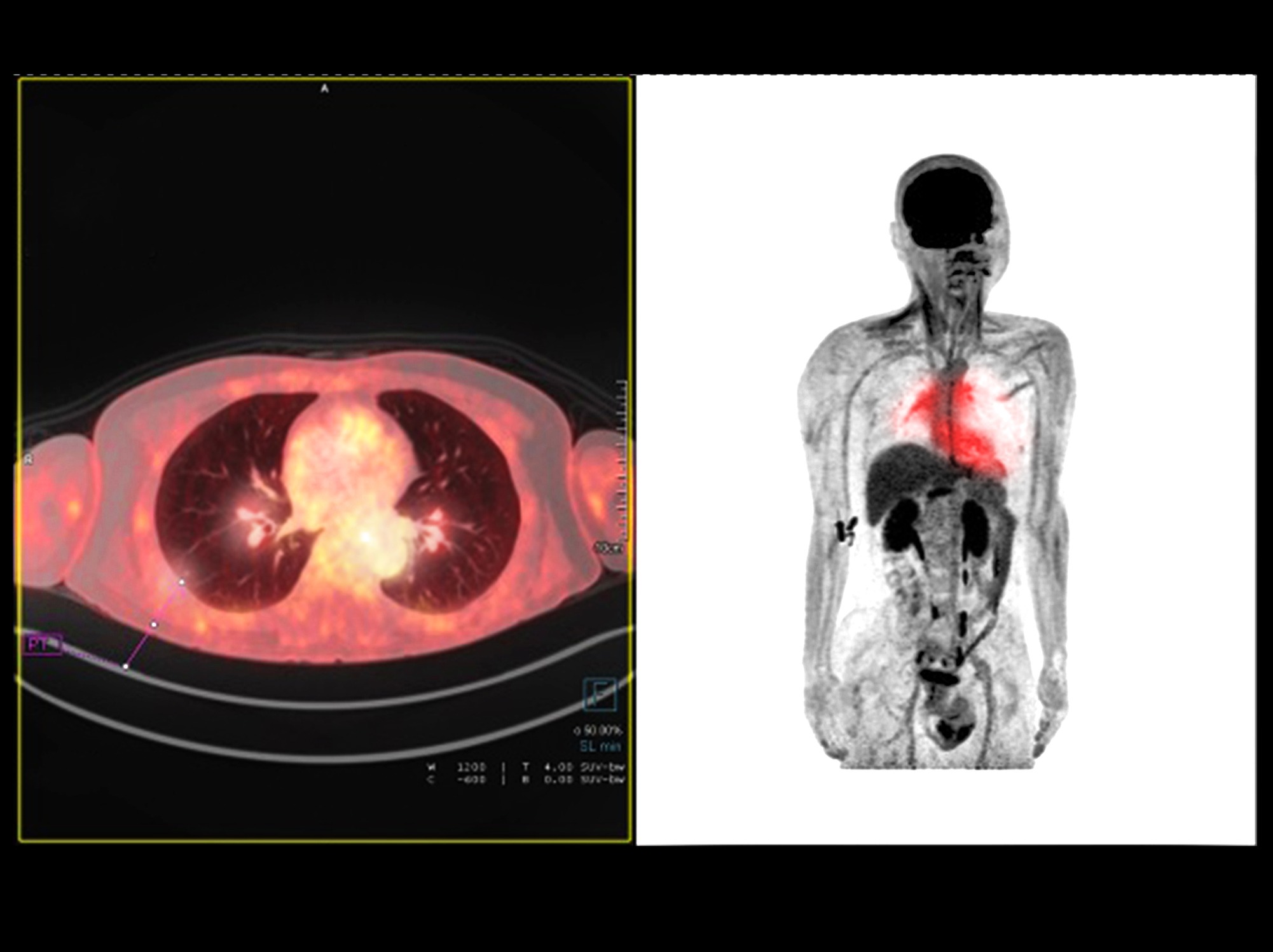
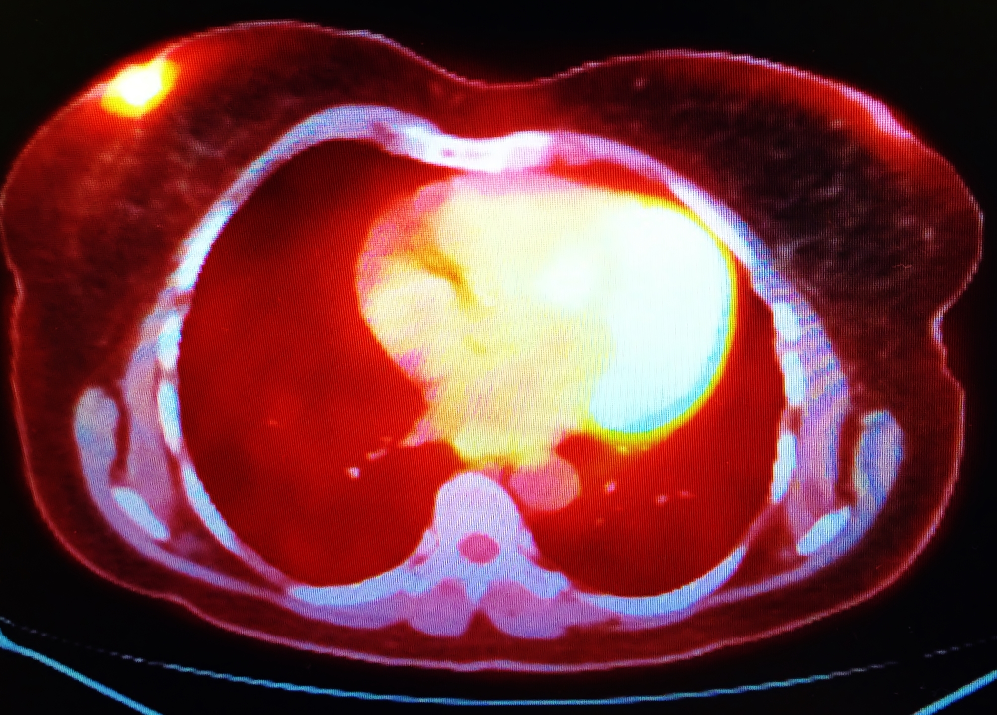
PET imaging is used in oncology, neurology and cardiology.
 By
By
Radiopharmaceuticals are used in nuclear medicine for the application of medical imaging and therapy.
 By
By
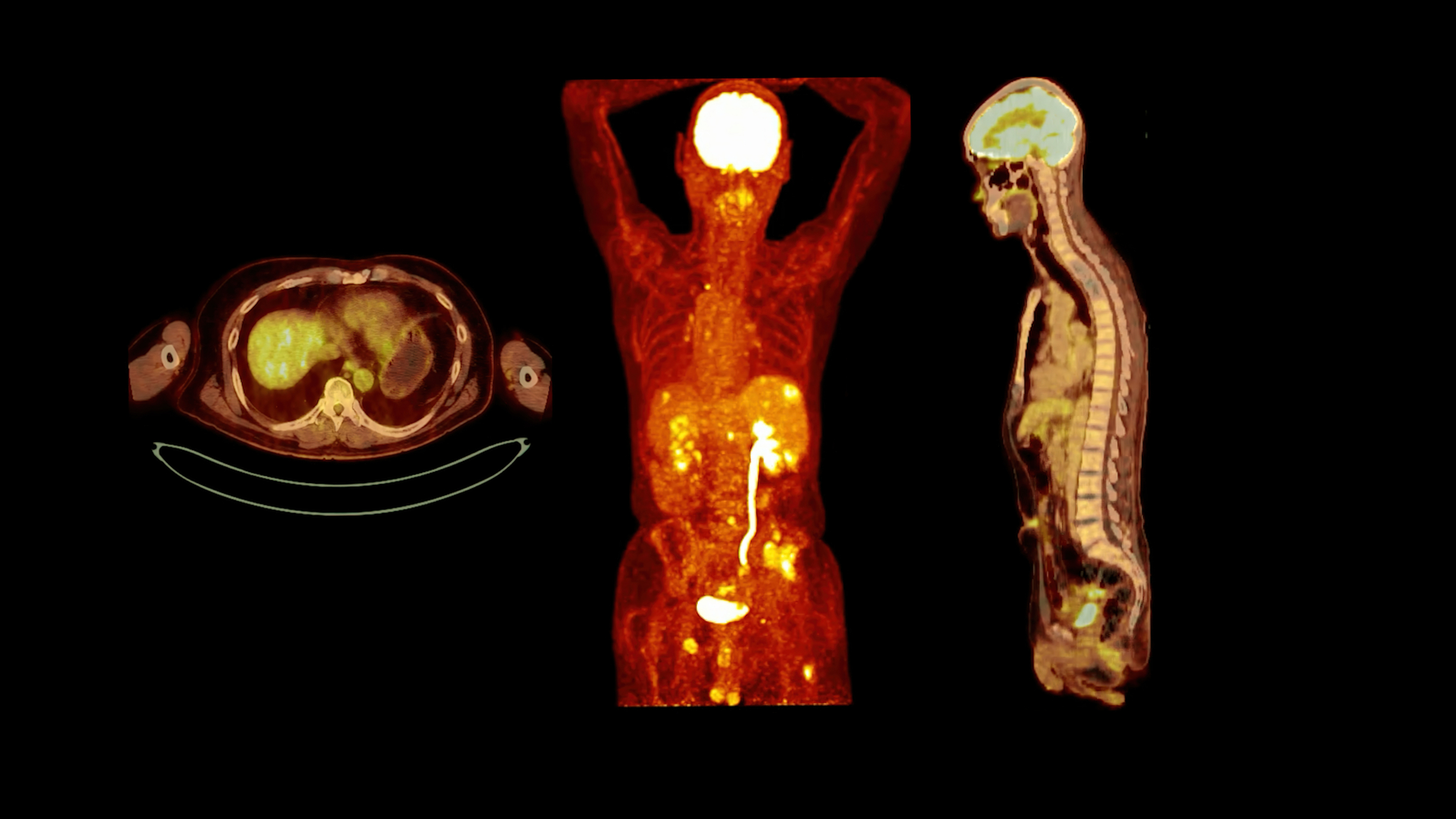
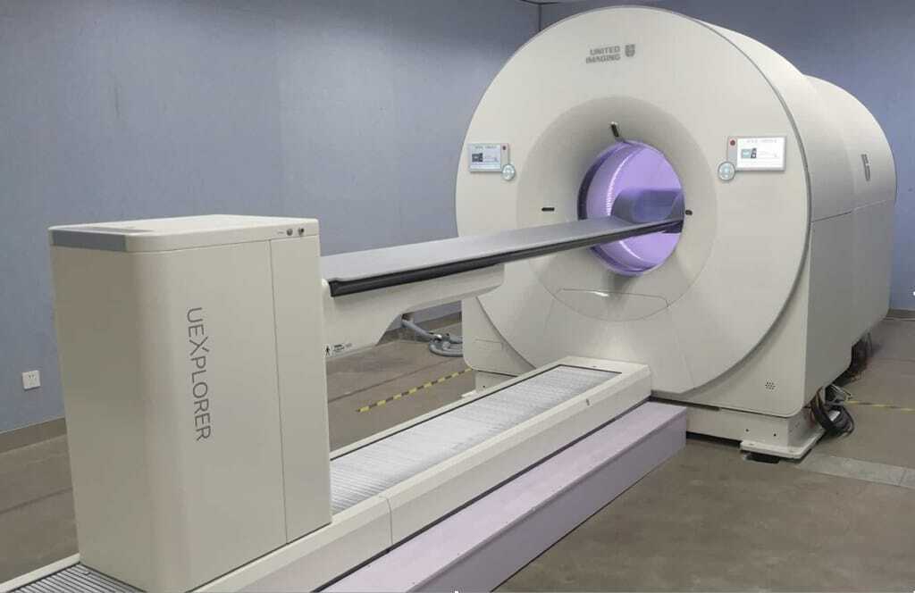
EXPLORER, the world’s first medical imaging scanner to produce a 3-D picture of the whole human body.
 By
By
The most commonly used medical radioisotope in diagnostic procedures is technetium-99m.
 By
By
The diagnostic breast imaging tool Positron Emission Mammography uses short-lived positron isotopes to detect breast cancer.
 By
By
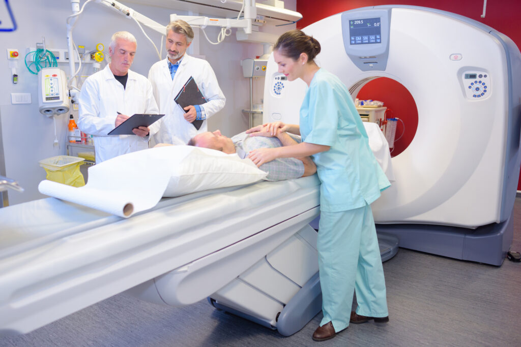
Nuclear Medicine Technologist is a specialised healthcare professional affiliated with a Nuclear Medicine Department. Image for illustration only. People depicted are models.
 By
By
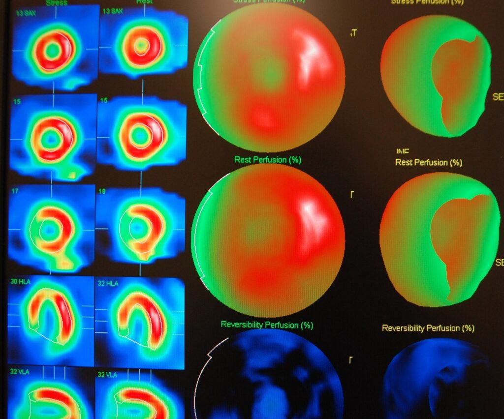
SPECT imaging is used to obtain a myocardial perfusion scan (SPECT scan) to investigate the function of the heart muscle.
 By
By
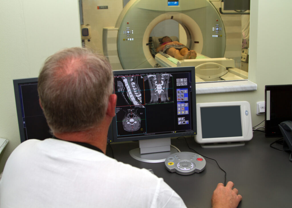
PET imaging (Positron Emission Tomography) is an established diagnostic imaging tool used in Nuclear Medicine.